


Description
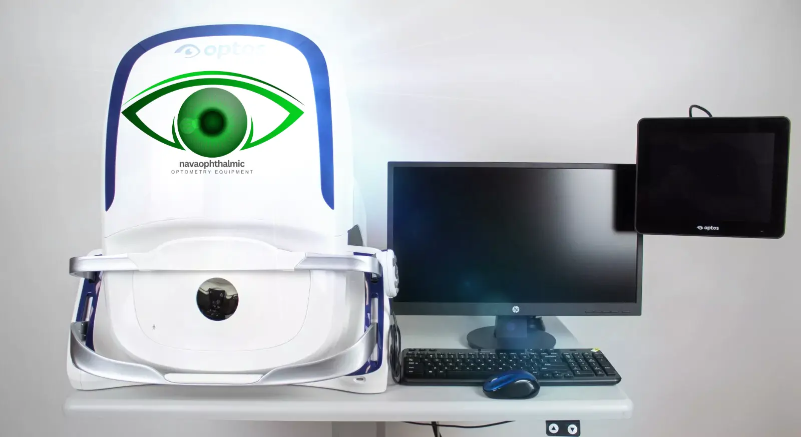
Engineers designed the Optos California for medical imaging. It comes in three models with multiple imaging modality options. California creates a clear 200° retinal image in under half a second. This technology is changing how we manage diseases like diabetic retinopathy, age-related macular degeneration, and uveitis.
If you had another fluorescein camera, then California will fit in the same area if much smaller. It also does autofluorescence, which is helpful for evaluating geographic atrophy
Dr. David M. Brown -Houstonretina.com
The system presents Optomap images in a consistent geometry that accurately represents anatomical features across the retina. Automatic image registration enables pixel-to-pixel comparisons of images across modalities and from visit to visit.
Optos has added new hardware and software in California. This helps doctors see more and find more eye problems. It also allows them to treat these issues better, which promotes patient health.
California’s advanced optical design gives clear images. It shows fine details when viewing the whole retina or zooming in on the macula, optic nerve head, or small issues.
Features and Benefits:
- Non-mydriatic imaging of the retina in less than ½ second saves time and helps improve clinic flow
- cSLO technology images through most cataracts and small pupils (2 mm)
- Multiple imaging methods are used. These include 3-in-1 color depth imaging, which shows color, red-free, and choroidal images in one. Autofluorescence also gives important clinical data. This data comes from the retinal surface to the choroid.
- Green laser autofluorescence shows macula and optic nerve head detail
- Image overlay tool facilitates comparison of images in different image modes and from visit to visit
- OptosAdvanceTM Image Management software streamlines image review and consultations
- DICOM compatible software supports compliance with the Code of Federal Regulations 1,2
- Distance (mm) and area (mm2) measurements provide objective assessment of change over time
- Stereo disc imaging allows assessment of the optic nerve to diagnose and follow the progression of glaucoma
- Auto-montage combines an optomap series into a single image appearing up to 220° (97%) of the retina.
- Motorized head and chin rest to more easily assist those patients requiring additional assistance during imaging
Technical Specifications:
- Image Modalities: optomap color and optomap plus (red and green laser):
- Color composite view
- Green laser view
- Red laser view
- optomap af (green laser): autofluorescence
- Resolution: optomap color: 20 μm, optomap plus, af : 14 μm
- Wavelengths: Red laser: 635 nm, Green laser: 532 nm
- Exposure Time: Less than 0.4 seconds
- Foot Print: Width: 550 mm / 22 inches
- Depth: 550 mm / 22 inches including chinrest
- Height: 608-632 mm / 24-25 inches
- Weight: 75 lbs
- Communication Protocol: DICOM Compatible

Effectively Cure more ocular conditions.
Optos introduces its newest ultra-widefield (UWF™) imaging apparatus, California, specifically for vitreoretinal specialists and ophthalmologists.
The compact desktop device offers better imaging features. It introduces a new imaging method called indocyanine green angiography, which is used to image the choroidal blood vessels. Optos claims it is important for diagnosing, managing, and treating certain conditions, including neovascular AMD and issues with choroidal neovascular membranes.
Why Optos California?
Optos has teamed up with California to create new hardware and software. This helps professionals see more and find more eye problems. As a result, they can treat more conditions and improve patient health. We’re committed to further strengthening our clinical evidence while demonstrating the importance of imaging the entire retina.
New proprietary optical hardware optimizes and maintains the resolution of their optomap images throughout the scan of the retina leading to more clarity at the far periphery.
Additionally, comparisons can make between different images or various dates by scrolling through all saved pictures.
The Optos California directional retinal imaging apparatus makes it possible for an immediate 200-degree perspective of a patient’s eye at a single scan, supplying a high-resolution picture in under half a second. Additionally, our advanced 3D OCT scanning instrument, the Topcon Triton, has updated software making glaucoma detection and decision making much easier and stronger
With optos California, an optometrist may generate over $9,000 – $15,000 a month. California Billed: $55 each picture for each single patients. Earning money every month with optos CaliforniaOptos California is available in three models:
California icg
California icg design for retinal specialists to maximize management of AMD, uveitic conditions along with other choroidal pathology; it offers the following imaging modalities and image viewing options.
California af
California af was made to facilitate retinal tests and document findings.
California fa
California fa was made to help identify and manage diabetic eye disease and other cardiovascular ailments.
Image Typeoptomap and optomap plus (red and green laser):
- Color composite
- green laser View
- red laser View
- optomap af (green laser): autofluorescence
- optomap fa (blue laser): fluorescein angiography
- optomap icg (infra-red): indocyanine green angiography
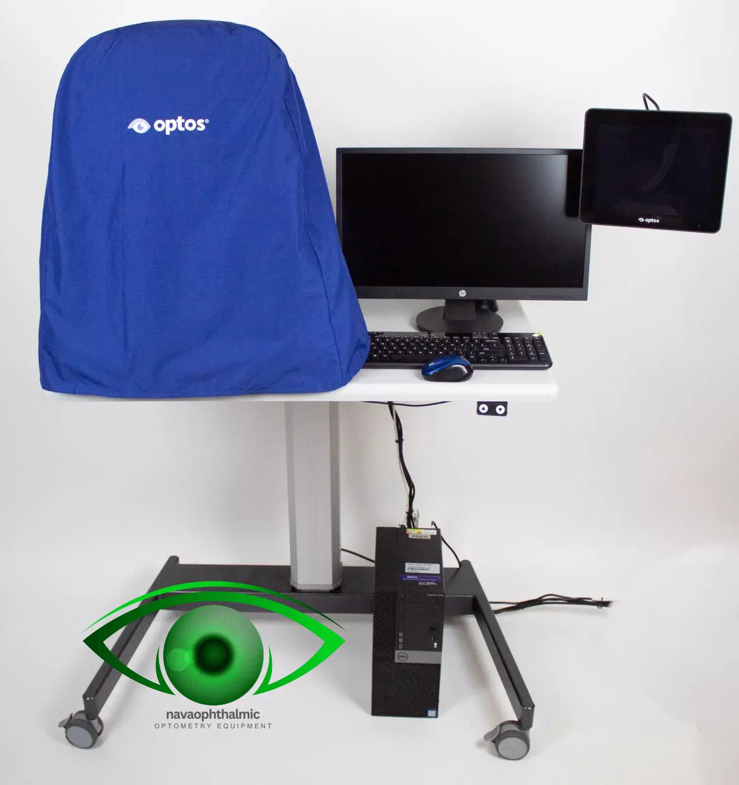
**Included with Optos California RG, AF:**
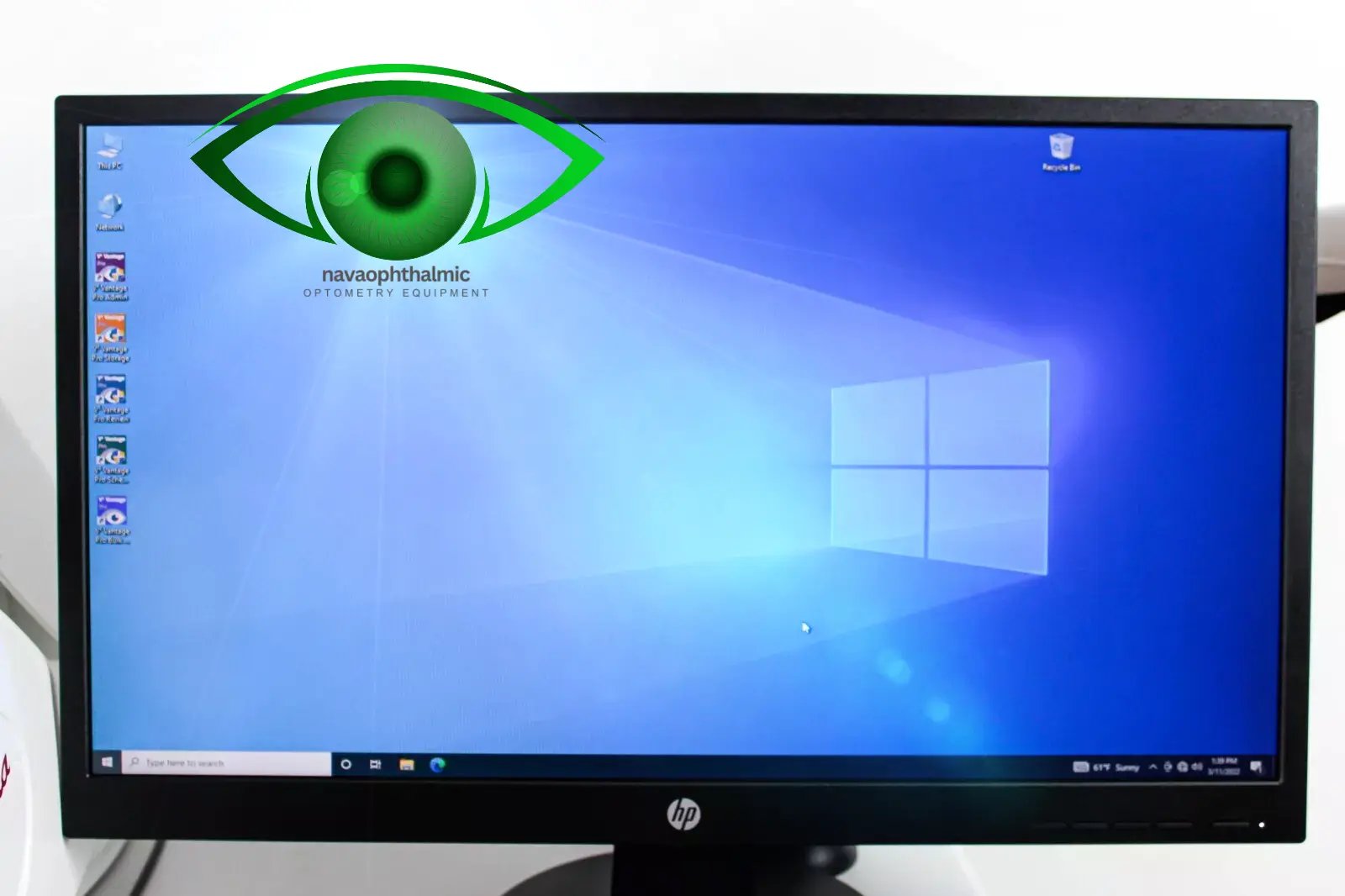
- – Windows 10 OS
- – New Dell Monitor
- – Power Table
- – Wireless Mouse + Keyboard


Reviews
There are no reviews yet.