Table of Contents
Understanding Optos Retinal Imaging Technology
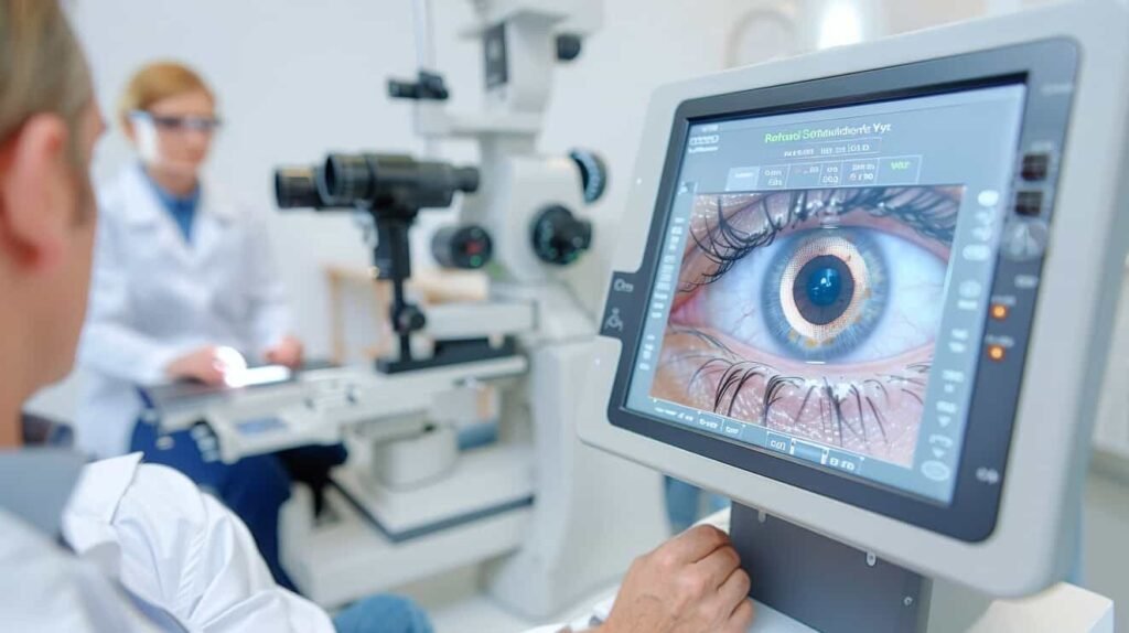
Retinal imaging is a critical facet of modern eye care. Providers can capture images of the interior structures of the eye quickly and with minimal discomfort to the patient.
Instant digital image delivery lets you examine the eye right away. This means patients do not have to wait for results. This, in turn, leads to earlier diagnosis of eye disease, which allows prompt treatment and better outcomes for patients.
Traditional retinal imaging has a limited field size. These devices can only capture 10 to 100 degrees of the retina in one image. This leads to incomplete imaging, and providers may miss potential pathologies.
Optos retinal imaging systems help reduce the chance of missing important details in retinal screenings by increasing the image field. The Optomap system is the first ultra-widefield imaging system. It offers a more complete retina image for a more comprehensive examination and accurate diagnostic findings.
Benefits of Optos Retinal Imaging
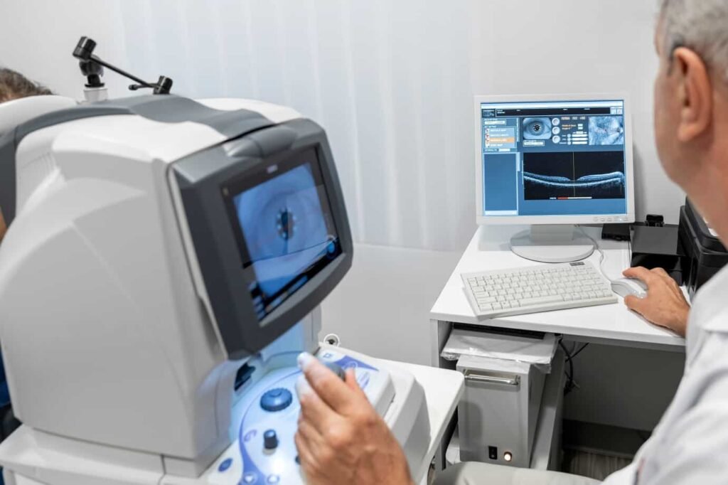
Optical coherence tomography (OCT) is a standard part of eye care. Eye care professionals use detailed OCT imaging to see the eye’s inner structures and check eye health.
Doctors use retinal screenings to diagnose common eye pathologies, including macular degeneration, diabetic retinopathy, and glaucoma. Finding problems like early signs of retinal detachment can help doctors act quickly to protect a patient’s vision.
Optos’ retinal imaging technology offers a broader imaging field than traditional retinal imaging devices. Optomap ultra-wide field retinal imaging can capture 82% or 200⁰ of the retina. With optomap auto-montage, eye care providers can capture up to 97% or 220⁰ of the retina in one image.
All Optos devices are non-mydriatic for improved patient comfort. There are no more lengthy waits in the office for dilation drops to take effect. Patients can return to normal activities immediately without the blurred vision and light sensitivity common with dilation.
Optos’ advanced eye imaging solution is very fast. The devices can take an image and create a 200-degree optomap in just half a second. The devices feature adjustable chin rests so patients are comfortable during imaging.
The Top Optos Retinal Imaging Devices for Eye Care Professionals
Optos offers a range of ultra-widefield (UWF™) non-mydriatic retinal imaging devices. All Optos ultra-widefield imaging devices capture high-resolution retinal images that allow professionals to discover, diagnose, document, and treat ocular pathology. The larger image range shows more of the retina, helping doctors find problems that may not appear with regular exams.
Providers can select the Optos retinal scanner that fits their needs. They can consider patient demographics, practice focus, and preferred image type.
1. Optos Daytona ($6,789.00)
Optos Daytona was designed to streamline eye screenings and improve the patient experience. The device produces a 200° single-capture optomap retinal image in less than half a second, making it ideal for practices that primarily see patients with good eye health.
Daytona is excellent for use on children and adults.
Clinical uses:
- Healthy refractive exams
- Diagnostic exams
- Disease monitoring
- Disease treatment
optomap Image Modalities
- Color rg
- Sensory red-free
- Choroidal
- Autofluorescence
Image Views
- Standard: 200⁰ Single Capture
- Auto-montage: Up to 220⁰
- Central Pole: Detailed view of the macula
- Stereo: Image pairing for optic disc and retinal evaluation
Software:
- OptosAdvanceTM Image Management software streamlines image review and consultations.
- The OptosAdvanceTM auto-montage tool combines many images to create a single 220° view that shows 97% of the retina.
- DICOM-compatible software supports compliance with the Code of Federal Regulations.
2. Optos California ($7,999.00)
The Optos California was developed with medical imaging in mind. It is highly effective for diagnosing, managing, and treating conditions such as Geographic Atrophy, Diabetic Retinopathy, AMD, and uveitis. The Optos California is available in various device configurations and image modality options to meet different practice needs.
Optos California makes a 200° retinal image in under half a second. This device is now a standard tool for retinal screening programs, and many doctors use it for patients at higher risk of eye problems.
Optos California is excellent for children, adults, and older adults.
Clinical uses:
- Healthy refractive exams
- Diagnostic exams
- Disease monitoring
- Disease treatment
- Medical procedures
Color rg
- Color rgb (available with rg/af/fa combined model)
- Sensory Red-free
- Choroidal
- Autofluorescence (green and blue*)(*feature may not be available in all regions)
- Fluorescein angiography
- Indocyanine green angiography
Image Views
- Standard: 200⁰ Single Capture
- Auto-montage: Up to 220⁰ (not available for fa or icg)
- Central Pole: Detailed view of the macula
- Stereo: Image pairing for optic disc and retinal evaluation
- 4-in-1 Color Depth Imaging
Software:
- OptosAdvanceTM Image Management software streamlines image review and consultations.
- The OptosAdvanceTM auto-montage tool combines many images to create a single 220° view that shows 97% of the retina.
- DICOM-compatible software supports compliance with the Code of Federal Regulations.
3. Optos MonacoPro
Optos MonacoPro offers a higher level of imaging with even greater efficiency than California or Daytona. MonacoPro captures five types of images for both eyes in just 90 seconds. It also provides 40° OCT views of retinal structures. The imaging device optimizes workflow while delivering superior patient care.
Optos MonacoPro is excellent for use on children, adults, and older adults.
Clinical uses:
- Healthy refractive exams
- Diagnostic exams
- Disease monitoring
optomap Image Modalities
- Color rg
- Sensory Red-free
- Choroidal
- Autofluorescence
- OCT
Image Views
- Standard: 200⁰ Single Shot
- Auto-montage: Up to 220⁰
- Central Pole: Detailed view of the macula
- Stereo: Image pairing for optic disc and retinal evaluation
- OCT: Cross-sectional imaging of ocular structures, including the fundus
Software
- OptosAdvanceTM Image Management software streamlines image review and consultations.
- A reference database (RDB) shows the OCT analysis results. It compares these results to 1%, 5%, 95%, and 99% of the RDB population.
- AreaAssist tool enables users to automatically measure continuous areas of matching color and adjust the sensitivity of the selected area.
- The OptosAdvanceTM auto-montage tool combines many images to create a single 220° view that shows 97% of the retina.
- DICOM-compatible software supports compliance with the Code of Federal Regulations.
4. Optos Silverstone
Optos Silverstone is the best tool for looking at the retina. The only ultra-widefield imaging device with built-in swept-source OCT is available. Silverstone creates a clear 200° single-capture retinal image in under half a second. It also allows for optomap-guided OCT scanning across the retina and into the far edges.
Optos Silverstone is excellent for use on children, adults, and older adults.
Clinical uses:
- Healthy refractive exams
- Diagnostic exams
- Disease monitoring
- Disease treatment
- Medical procedures
optomap Image Modalities
- Color rg
- Sensory Red-free
- Choroidal
- Autofluorescence
- Fluorescein angiography
- Indocyanine green angiography
- OCT
Image Views
- Standard: 200⁰ Single Capture
- Auto-montage: Up to 220⁰ (not available for fa or icg)
- Central Pole: Detailed view of the macula
- Stereo: Image pairing for optic disc and retinal evaluation
- OCT: Up to 23mm line scan and swept-source functionality
- Automatic rescan function offers precise follow-up scanning
Software
- OptosAdvanceTM Image Management software streamlines image review and consultations.
- The OptosAdvanceTM auto-montage tool combines many images to create a single 220° view that shows 97% of the retina.
- DICOM-compatible software supports compliance with the Code of Federal Regulations.
Which Optos Device is Right for Your Practice?
All the Optos devices offer UWF imaging with multiple image modalities. All models improve your diagnostic capabilities for better patient outcomes.
Choosing the best retinal imaging device for your practice depends on various factors. Elements to consider include:
- Patient population
- Appointment volume
- Facility space
- Budget
You can also consider what eye care needs you are meeting for your patients. To correct vision for patients with good eye health, the Optos Daytona may help you.
If you screen many patients with risk factors for eye disease, the Optos California model may be the best choice. Medical professionals can use the Optos California and Optos Silverstone for procedures. The Optos MonacoPro is excellent for treating eye conditions.
Why Choose Optos Retinal Imaging for Your Optometry Practice?
Optos retinal imaging can benefit any eye care practice. Regardless of patient age and health status, retinal images are an integral tool for eye health. The speed and ease of Optos devices make them easy to integrate into your appointment workflow. You can accommodate more patients without sacrificing screening quality or patient experience.
Optos is safe for patients of all ages, including pediatric and geriatric populations. Since there is no need for dyes or dilution, it is easy for children to cooperate with the process.
Images are available within moments so that doctors can assess them during the appointment. They can also be saved in patient electronic health records, allowing for comparison over time.
The Optos system is excellent for managing chronic eye conditions like diabetic retinopathy and glaucoma.
New materials are added often, allowing providers to participate in on-demand learning. These opportunities help them better serve their patients.
For more information on Optos UWF retinal imaging devices, visit the Optos website. You can also contact Nava Ophthalmic to learn which device is best for your eye care practice. Learn about the benefits of optomap imaging today.

John Berdahl, MD
Meet Dr. Berdahl
Dr. Berdahl is most motivated by the trust his patients place in him during their moments of vulnerability.
That patient trust has, first and foremost, driven him to become an accomplished surgeon. However, as he meets patient needs and learns more about the problems they face, he’s had several opportunities to stretch his skills as an inventor and problem-solver. He co-invented the MKO melt, an innovation used in our Sioux Falls clinic that provides sedation during cataract surgery without the use of an IV or opioids, and developed Interfeen, a rare disease drug that helps with ocular conditions. He created astigmatismfix.com, a resource that has helped tens of thousands of surgeons eliminate residual astigmatism after cataract surgery, and he co-founded ExpertOpinion.MD, a site where patients can request medical opinions from authentic world experts. He also is the founder of Balance Ophthalmics, the first non-surgical, non-pharmacologic way to lower eye pressure for glaucoma treatment, which was FDA approved in 2024.
Interests & Added Expertise
Dr. Berdahl is equipped to employ the most innovative and tested techniques available to effectively treat most diseases of the anterior segment (front part of the eye). He is exceptionally skilled at diagnosing the best treatment for varying stages of glaucoma, corneal diseases, and cataracts. He is also a meticulous refractive surgeon.
To advance the technologies available to our patients, Dr. Berdahl collaborates with numerous ophthalmology companies as a consultant. However, to minimize potential bias, all consulting fees are donated to charity.
Education
- Hills-Beaver Creek High School, Hills, MN
- Augustana College, Sioux Falls, SD
- Mayo Medical School, Rochester, MN
- Mayo Clinic Internship, Scottsdale, AZ
- Duke University, Durham, NC
- Minnesota Eye Consultants Fellowship (Minneapolis, MN)