Table of Contents
Retinal imaging plays a critical role in modern eye care. From detecting early signs of diabetic retinopathy to monitoring macular degeneration, high-quality fundus imaging has become essential for delivering exceptional patient outcomes.
Between rising patient demand and rapid advances in diagnostic technology, eye care professionals need reliable solutions that provide precise, wide-field retinal images quickly.
Enter the ZEISS Fundus Camera lineup, designed for:
- Optometrists
- Ophthalmologists
- Eye care clinics
These devices redefine what’s possible in retinal diagnostics. It doesn’t matter whether you’re running a solo practice or a large medical clinic. They elevate the standard of care by offering:
- Robust functionality
- User-friendly interfaces
- Powerful imaging features
Are you seeking to enhance patient outcomes, clinic efficiency, and diagnostic accuracy? The ZEISS Fundus Camera is your answer.
Please keep reading to explore how it can transform your practice today.
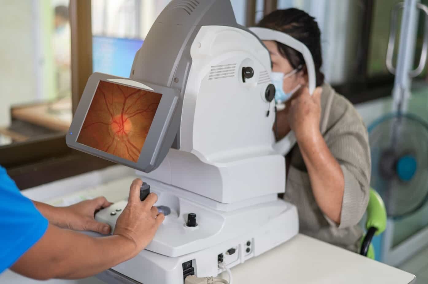
What Is a Fundus Camera?
A fundus camera takes clear pictures of the inside of the eye. It gives crucial visual information for doctors to diagnose conditions. It focuses specifically on key retinal structures, including the following:
- Retina
- Optic disc
- Macula
- Posterior pole
These images allow eye care professionals to examine clearly:
- Fine details of blood vessels
- Nerve fiber layers
- Central retinal zones
Unlike traditional ophthalmoscopes, fundus cameras offer wide-angle views and digital documentation. This enables more thorough and consistent assessments over time. In modern optometry and ophthalmology, they are indispensable tools for diagnosing and monitoring ocular diseases with precision.
It enhances patient care and outcomes by:
- Detecting early signs of glaucoma, diabetic retinopathy, macular degeneration, and other vision-threatening conditions
- Documenting disease progression through serial imaging
- Providing visual evidence to support treatment plans
Traditional ophthalmoscopes offer limited views and often require significant expertise and time to interpret findings. A modern fundus camera captures wide, detailed images with consistency, enabling faster and more accurate diagnosis.
These digital systems create a visual record. This record can be stored, compared, and shared for long-term care planning.
Retinal imaging technology for optometrists and ophthalmologists is a clinical necessity, not a luxury. With the right camera, clinicians can:
- Diagnose earlier
- Explain conditions clearly to patients
- Track improvements or progression over time
The use of digital fundus imaging does the following:
- Elevates clinical documentation
- Aids in insurance approvals
- Supports patient understanding and adherence to treatment
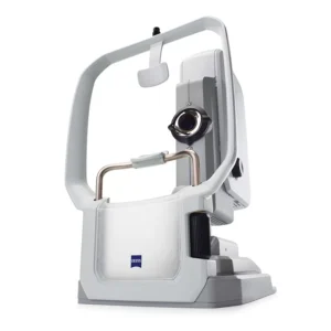
Why Choose the ZEISS Fundus Camera?
ZEISS, a global leader in optical and medical technology, has a long-standing reputation for precision, craftsmanship, consistent reliability, and ongoing innovation. For over a hundred years, ZEISS has led advancements in diagnostic imaging. They set high standards for optical quality in medicine.
Their fundus cameras reflect this legacy by offering:
- Uncompromising quality
- Advanced imaging capabilities
- Intuitive design that meets the needs of both optometrists and ophthalmologists
From groundbreaking wide-field imaging to seamless software integration, ZEISS devices consistently exceed the expectations of clinicians worldwide. Their commitment to continual improvement and clinical excellence makes ZEISS a trusted name in every modern eye care environment.
What sets ZEISS fundus Cameras apart:
- True color ultra-widefield imaging for comprehensive views of the retina in its natural state
- Multimodal imaging including color, red-free, and autofluorescence modes
- Intelligent autofocus and alignment simplify acquisition for staff
- Seamless EMR integration enables smoother workflow and faster charting
A fundus camera for ophthalmologists can provide:
- Accuracy
- Speed
- Ease of use
- Integration into a busy clinical workflow
These professionals rely on advanced diagnostic tools to assess complex retinal conditions with precision and efficiency. ZEISS exceeds expectations in all areas by delivering:
- Exceptional image resolution
- Rapid capture times
- Intuitive user interfaces
This reduces training time and enhances staff productivity. ZEISS fundus cameras are reliable and perform well in both busy eye care centers and specialty retina clinics.
Additional benefits include the following:
- Increased clinical productivity with fast acquisition and minimal patient prep
- Minimized motion artifacts with advanced autofocus
- Enabling confident diagnosis with high-definition imaging
- Easily sharing visual results with patients for better communication
ZEISS devices also support remote consultation models, allowing images to be reviewed by specialists off-site. This ability is extremely useful in practices with multiple locations. Quick input from a retinal specialist also holds great importance.
You can securely transfer high-quality images, enabling collaborative diagnosis and faster treatment planning. As telemedicine gains importance, ZEISS imaging systems would allow practices to reach a wider range of patients. They can provide expert care no matter the distance.
This flexibility proves especially useful in rural communities and underserved populations where specialists may have limited access. By facilitating efficient collaboration and referral processes, ZEISS enhances continuity of care and improves outcomes across diverse patient settings.
Top ZEISS Fundus Cameras in the Market
ZEISS Ophthalmic offers a versatile range of fundus cameras designed to meet the specific demands of various clinical environments. ZEISS offers a range of solutions for all types of practices and diagnostic needs. They have compact, portable models and advanced imaging systems with integrated OCT.
This section explores the top ZEISS fundus Cameras available. It breaks down their key features. It also looks into clinical advantages and best use cases.
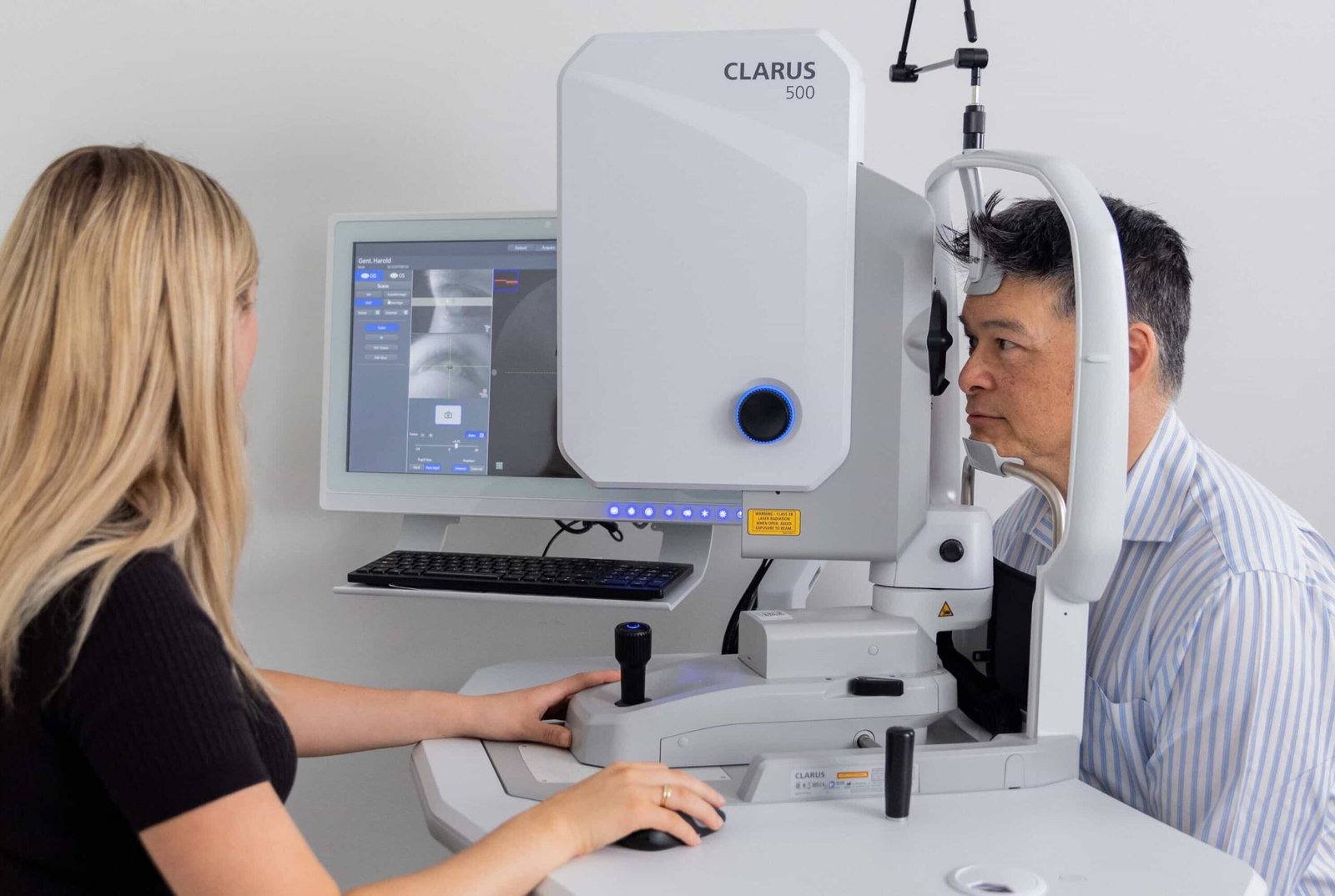
1. ZEISS CLARUS 500
The ZEISS CLARUS 500 combines widefield coverage with unmatched true color fidelity and advanced imaging performance. It features a 133° field of view in a single capture.
You can expand this to 200° with montage. This provides a full view of both the central and side areas of the retina. Its optics create images that closely match the natural colors of the retina. This enhances diagnostic accuracy for subtle vascular or pigmentary changes.
The CLARUS 500 supports non-mydriatic imaging, allowing faster exams without the need for dilation. It includes red-free and autofluorescence modes for additional diagnostic flexibility.
Key features:
- High-resolution true color imaging
- Non-mydriatic operation
- Red-free and autofluorescence modes
- Dual-eye capture for stereo imaging
Clinical advantages:
- Detect fine vascular changes and early pathology
- Reduce light exposure for patient comfort
- Perform fast, reliable diabetic retinopathy screening
- Improve care for AMD and retinal vascular disease
Best for:
- Optometry clinics offering diabetic eye exams
- Practices focused on retinal disease detection
- Group practices seeking scalable imaging solutions
2. ZEISS CLARUS 700 ($7,779.00)
The ZEISS CLARUS 700 builds upon the 500’s capabilities by incorporating multimodal functionality, providing clinicians with greater diagnostic flexibility. It enhances retinal imaging with ultra-widefield capture up to 200° in a single shot. This allows visualization of peripheral retinal pathology in stunning true color.
This model is made for detailed retinal analysis. It supports advanced imaging needs with different imaging modes and easy data comparison tools.

Key features:
- True color, autofluorescence, and infrared imaging
- Integrated optical coherence tomography (OCT)
- QuickView image comparison for disease tracking
- SmartTouch interface for ease of use
Clinical advantages:
- Combines structural and functional imaging in one unit
- Enables precise monitoring of geographic atrophy, glaucoma, and macular edema
- Improves clinical efficiency and patient throughput
Ideal for:
- Ophthalmology clinics managing advanced retinal pathology
- Specialists conducting OCT-guided diagnostics
- Large practices wanting to consolidate equipment
3. ZEISS VISUSCOUT 100
Compact and portable, the ZEISS VISUSCOUT 100 is ideal for point-of-care imaging and mobile eye care. It bridges the gap between advanced diagnostic capability and portability. This makes it a critical asset for clinics seeking flexibility.
The device allows clinicians to bring high-quality retinal imaging to patients in remote, underserved, or immobile environments. Its lightweight, wireless design removes the need for big equipment setups. It does not compromise on image quality or data transfer reliability.

Key features:
- Weighs less than one pound
- Captures 40° retinal field
- Rechargeable battery with 4-hour runtime
- Wi-Fi image transfer to PC or EMR
Clinical advantages:
- Enables outreach services in remote or underserved areas
- Improves workflow for optometrists performing off-site exams
- Ideal for elderly and pediatric patients with mobility issues
Best for:
- Mobile vision units and public health programs
- Nursing home and in-home eye exams
- Budget-conscious practices that want mobility
How ZEISS Fundus Cameras Improve Patient Care
Early detection saves vision. The ZEISS Fundus Camera lineup empowers providers to detect, document, and treat eye diseases before irreversible damage occurs.
ZEISS systems take clear, high-resolution images in natural color. This helps doctors detect small changes in the retina that they might otherwise miss. This proactive approach enables:
- Timely interventions
- Improved long-term outcomes
- Greater peace of mind for both doctors and patients
Clinical impact:
- Identify microaneurysms in diabetic retinopathy
- Spot optic nerve head changes for glaucoma diagnosis
- Monitor the progression of age-related macular degeneration
High-definition imaging supports evidence-based care plans and improves follow-up compliance. Patients understand their diagnosis when shown clear visuals.
Visual confirmation helps demystify complex conditions and builds patient confidence in the prescribed treatment path
These images also act as motivation. They encourage patients to follow care plans and come back for regular check-ups.
Ultimately, clear and engaging visuals foster stronger communication between providers and patients, leading to more effective care outcomes.
Additional advantages include:
- Reduced imaging time improves patient satisfaction
- Enhanced documentation supports insurance billing and referrals
- Visual proof increases acceptance of recommended treatments
Buying Guide: Choosing the Right ZEISS Fundus Camera
Selecting the best fundus imaging solutions for your clinic depends on various factors. Your decision should be based on:
- Clinic size
- Patient demographic
- Diagnostic needs
Different models offer varying degrees of:
- Mobility
- Field of view
- Multimodal capabilities
Evaluating these features alongside your current workflow ensures a seamless fit that supports both clinical excellence and operational efficiency.
Practice size:
- Solo or small clinics: ZEISS VISUSCOUT 100
- Mid-size clinics: ZEISS CLARUS 500
- Large practices or hospitals: ZEISS CLARUS 700
Patient volume:
- High-volume screening clinics need fast, widefield capture
- Specialty clinics require multimodal features and OCT
Clinical focus:
- Primary care optometry: CLARUS 500 or VISUSCOUT 100
- Retina subspecialty: CLARUS 700 with OCT
Budget considerations:
- VISUSCOUT 100 offers the most affordable entry point
- CLARUS 700 provides the most comprehensive feature set
EMR compatibility:
- All models integrate with leading EMR platforms for easy data transfer
How to Purchase ZEISS Fundus Cameras From Nava Ophthalmic
Nava Ophthalmic is a top company that distributes fundus imaging solutions and optometry equipment. They serve clinics, hospitals, and eye care professionals worldwide. They are committed to delivering only the highest quality ophthalmic products.
Nava ensures that every clinic has access to the tools needed for top-tier diagnostic care. Our extensive inventory includes advanced imaging systems, such as those from ZEISS, backed by thorough product knowledge and responsive customer support.
Eye care professionals trust Nava to provide innovative, reliable solutions that align with clinical goals and enhance patient outcomes. Nava offers:
- Expert product consultations
- Fast shipping through optimized logistics
- Certified installation and user training
- Long-term technical support and warranty services
Nava Ophthalmic is not only a supplier; it is a strategic partner. The team is dedicated to helping clinics thrive and provide patients with the best available equipment. Our catalog includes only proven, reliable ophthalmic tools that meet rigorous standards.
Ask about exclusive ZEISS bundle deals and seasonal financing options.
Frequently Asked Questions
Are you still wondering whether a ZEISS Fundus Camera from Nava Ophthalmic is the right choice? Answering common FAQs can help you make an informed decision.
What makes ZEISS Fundus cameras different from other brands?
ZEISS provides true color, widefield retinal imaging, user-friendly interfaces, and seamless EMR integration. They have built their reputation on over a century of precision optics.
Can ZEISS Fundus Cameras Integrate With EMR Systems?
All ZEISS models work with major EMR software platforms. This enables efficient storage of images and data.
What Is the Warranty and Support Provided by Nava Ophthalmic?
Purchases from Nava Ophthalmic include:
- Installation
- Training
- Technical support
- Warranty coverage
Extended service options are also available.
Are There Training Resources Available for New Users?
Nava offers onboarding support, user guides, and live training. This is to ensure you and your team maximize your ZEISS camera features.
How Long Does It Take to Receive My ZEISS Fundus Camera?
Most models ship quickly through Nava Ophthalmic’s distribution centers. Estimated delivery times vary by location and stock.
Can I Finance the Purchase of a ZEISS Fundus Camera?
Flexible payment and financing plans are available. Contact Nava Ophthalmic for current financing options.
Upgrade Patient Care With the ZEISS Fundus Camera Today
ZEISS Fundus Cameras lead the way in retinal imaging for optometrists, ophthalmologists, and vision clinics. They provide speed, accuracy, and confidence for every patient exam. This is true whether you are checking for changes in diabetes or managing long-term eye disease.
Each model provides a tailored experience tailored to your clinic’s size, goals, and patient population. In the competitive world of eye care, having access to leading eye care devices is essential for excellence.
Shop ZEISS Fundus Cameras from Nava Ophthalmic today and redefine the standard of retinal imaging in your practice.