Description
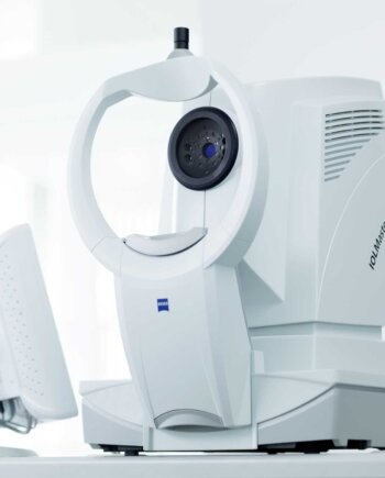
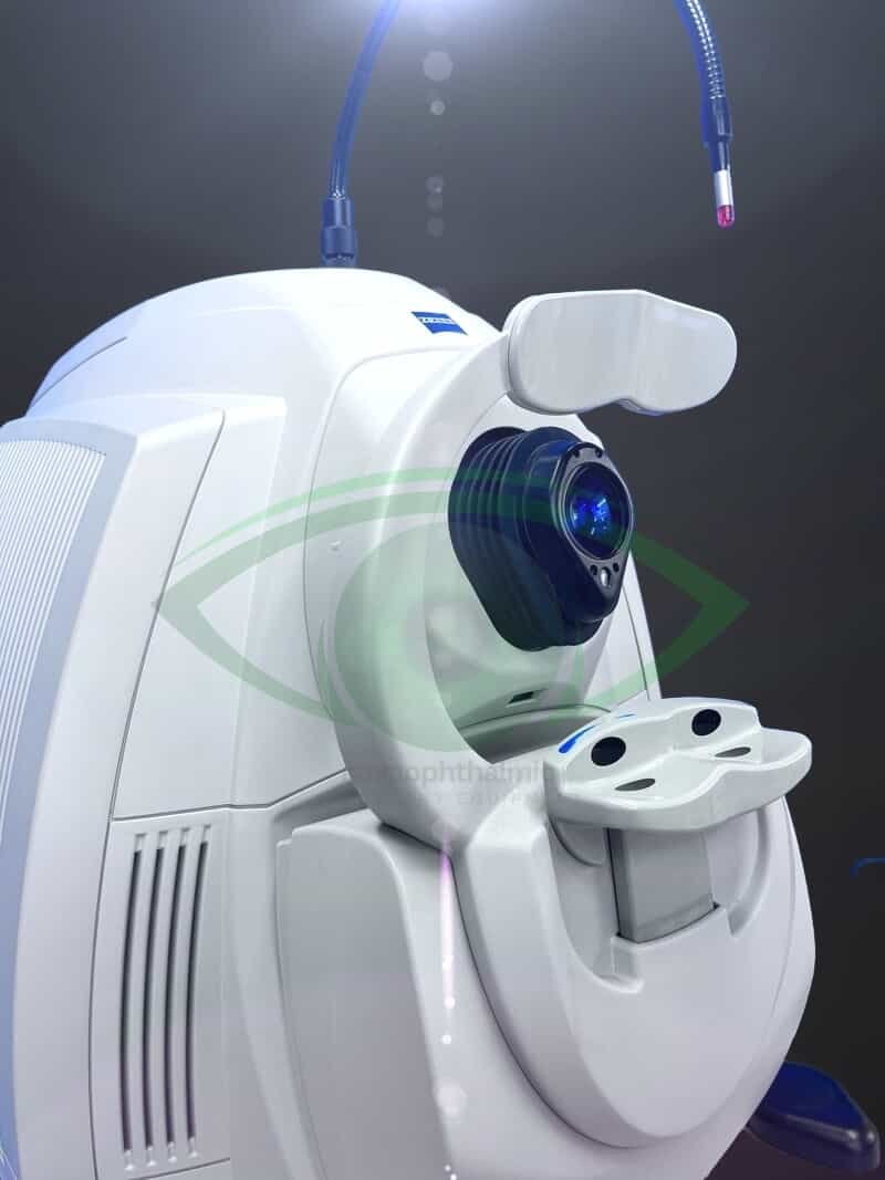
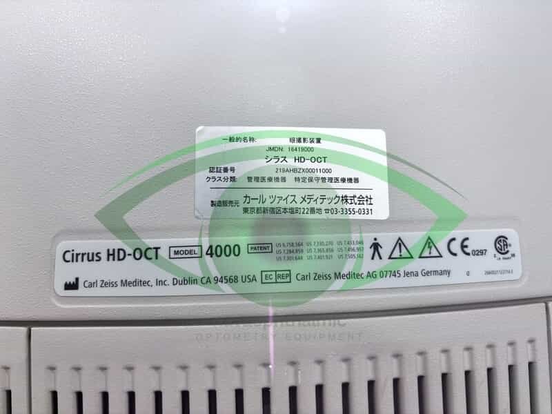
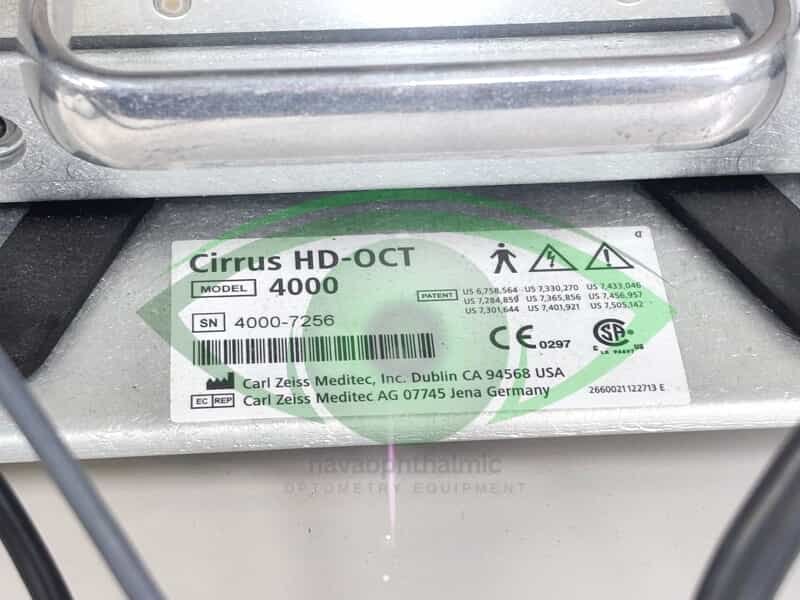
CARL ZEISS Cirrus HD-OCT 4000 analyzes glaucoma and retina with modern integrated design, ease of use, and a small footprint.
With the smaller budget designed Cirrus HD-OCT 4000 focused on the essential core OCT functionality.CARL ZEISS Cirrus HD-OCT Have two models, Model 400 and Model 4000, which are capable of anterior segment imaging and offer the same package of glaucoma and retina analyses
Cirrus HD-OCT 4000 allows visualization and analysis of a patient’s condition and captures a tightly packed, detail-rich cube of data in just seconds making this unit the best-selling spectral domain OCT system
Computer
• High-performance multi-core processor
• Internal storage: > 80,000 scans
• CD-RW, DVD-ROM drive
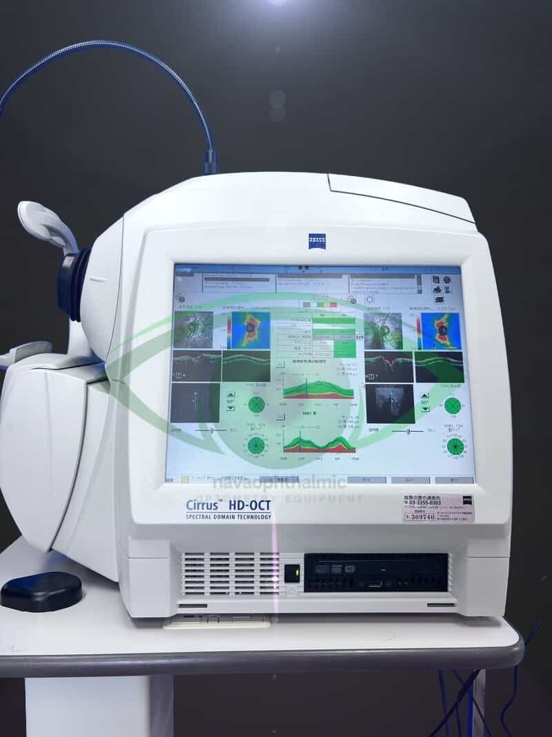
• Integrated 15″ color flat panel display
CARL ZEISS Cirrus HD-OCT 4000 FEATURES
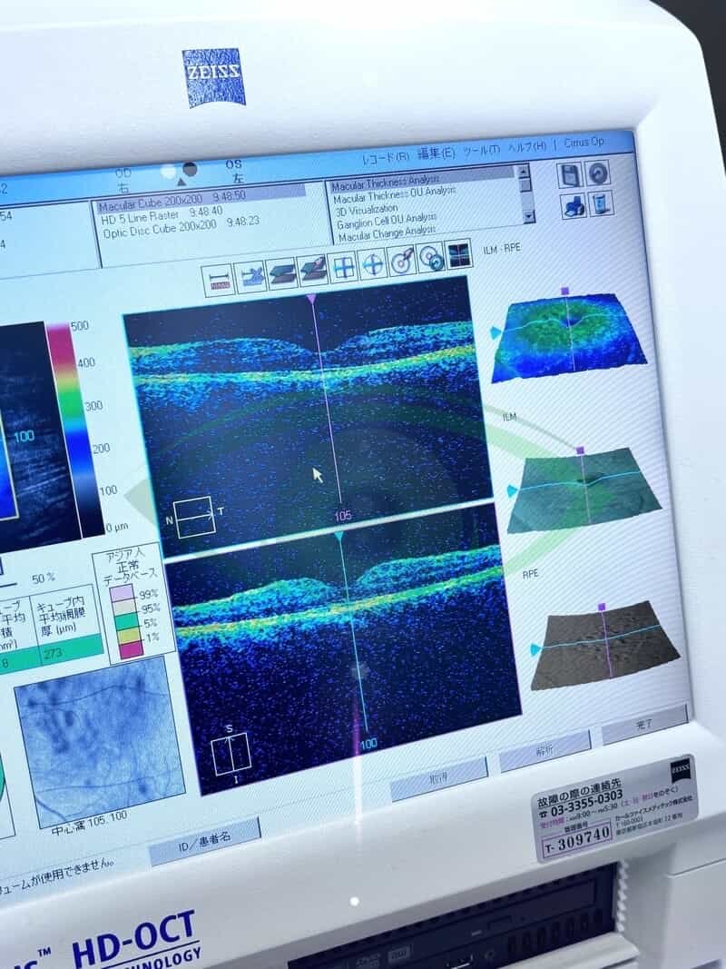
- Unprecedented Visualization of Anatomical Details
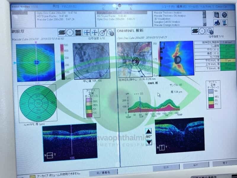
- HD Layer Maps
- HD Enhanced Raster Scan
- Cirrus HD-OCT Scan Engine
- Correlation Between OCT Scan and Fundus Image
- Superior Image Data
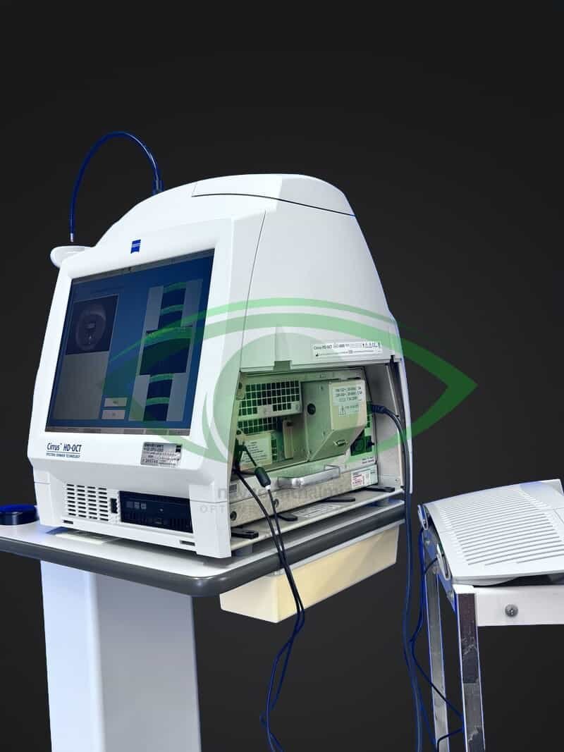
- Designed for efficiency
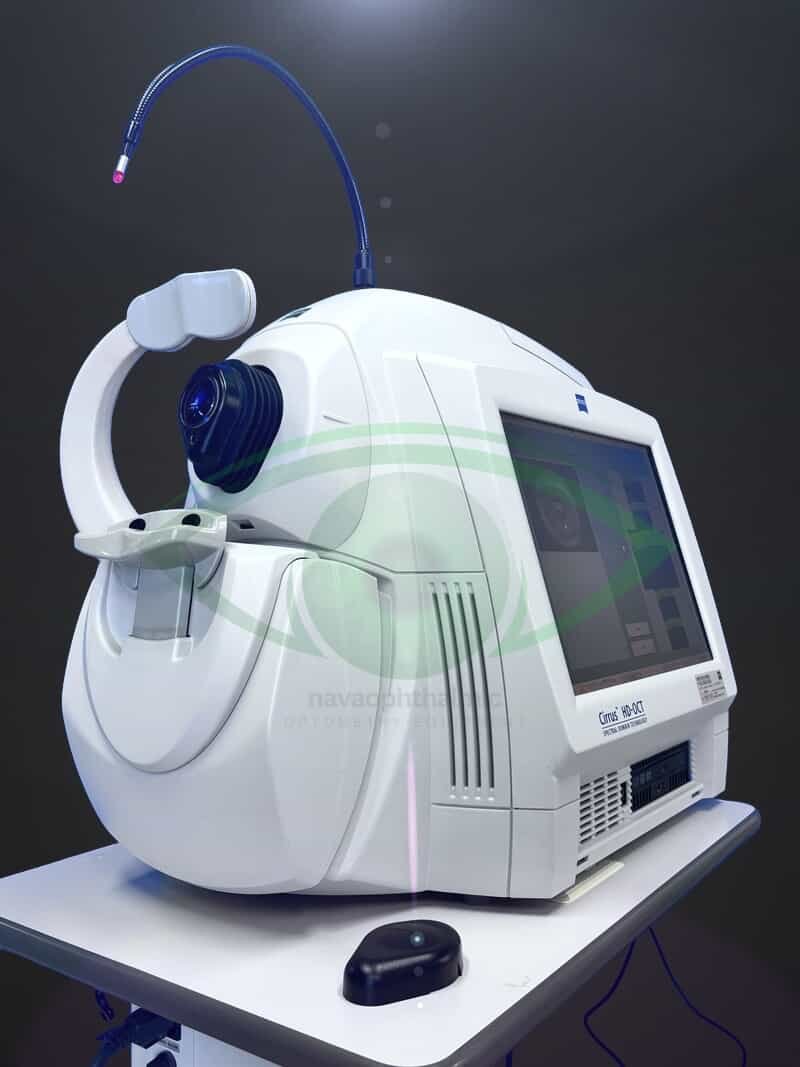
- Small footprint and integrated design are ideal for crowded or busy practice
- 90 degree orientation facilitates observation of patient throughout exam
- Advanced optics aid in the examination of patients with cataracts. Dilation is not required even for pupils as small as 2.5 mm
- Mouse Driven Alignment™ delivers superior image capture and analysis in just a few clicks, resulting in reduced chair time for the patient
- Auto Patient Recall™ assures patient position and instrument setting are repeated from previous visit
SPECIFICATIONS
Technical data
- Axial resolution: 5 μm (in tissue)
- Transverse resolution: 15 μm (in tissue)
- Scan speed: 27,000 A-scans per second
- A-scan depth: 2.0 mm (in tissue), 1024 points
- Optical source: superluminescent diode (SLD), 840 nm
Fundus Imaging
- Line scanning ophthalmoscope (LSO)
- Live during scanning
- Transverse resolution: 25 μm (in tissue)
- Optical source: superluminescent diode (SLD), 750 nm
- Field of view: 36° x 30°
Scan Patterns
- Macular Cube 200 x 200 Combo: 200 horizontal scan lines comprised of 200 A-scans
- Macular Cube 512 x 128 Combo: 128 horizontal scan lines comprised of 512 A-scans
- 5 Line Raster: 4096 A-scans per B-Scan (adjustable length, spacing and orientation)
Focus Adjustment Range
- –20D to +20D (diopters)
Fixation
- Internal and external
Pupil Size Requirement
- ≤ 2.0 mm (≥ 3.0 mm optimal for LSO)
Computer
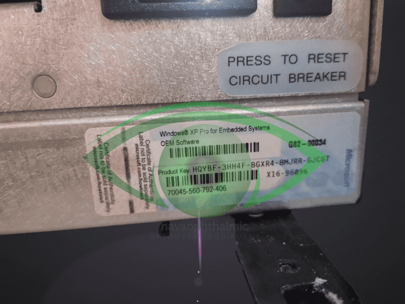
- Windows® X 7 Ultimate
- High-performance multi-core processor
- Internal storage: > 80,000 scans
- CD-RW, DVD-ROM drive
- Integrated 15” color flat panel display
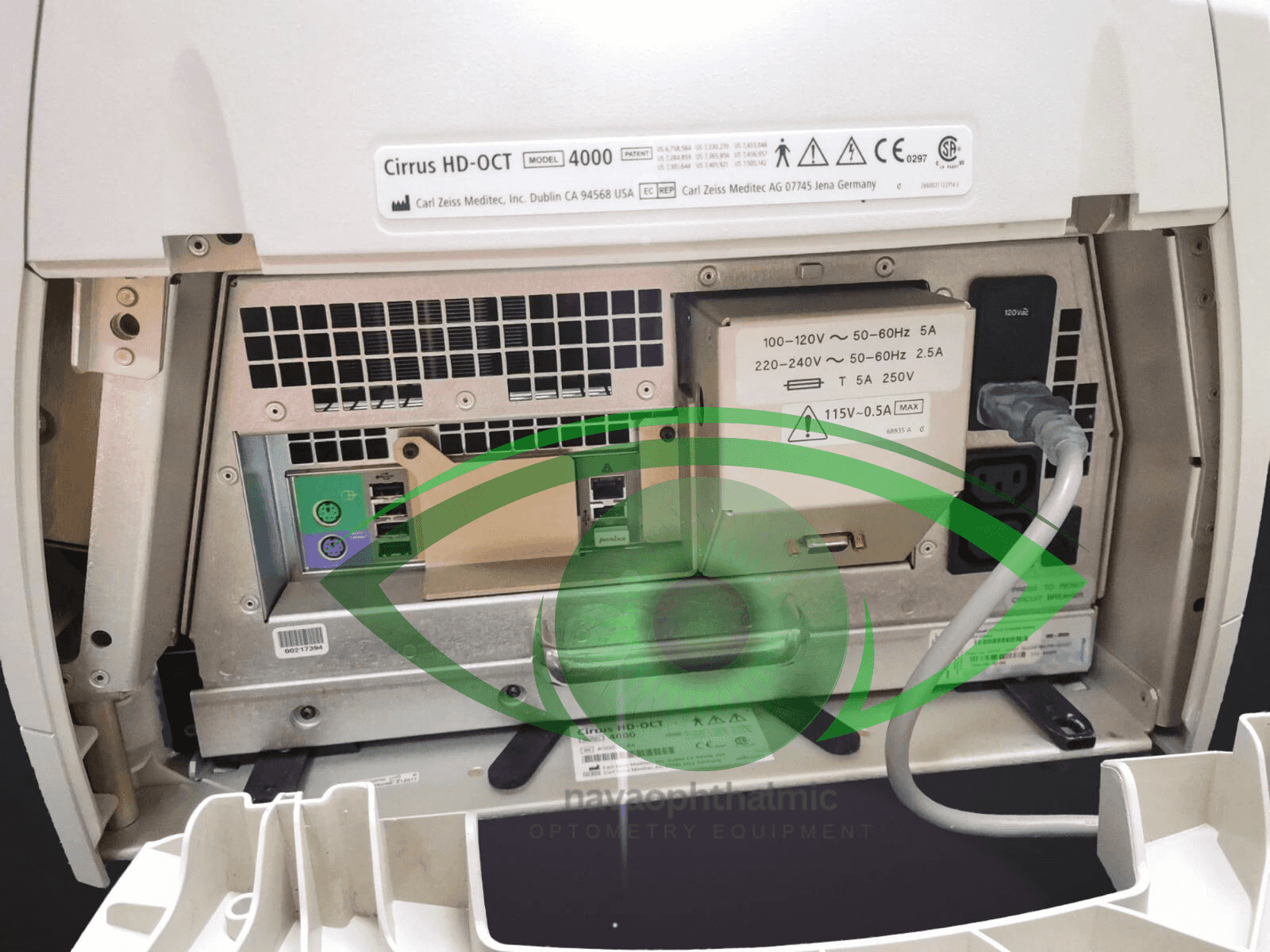
Electrical
- 100–120V~, 50/60Hz, 5A 220–240V~, 50/60Hz, 2.5A
INCLUDES
- -Manual
- -Additional Cords / Cables
- -Additional Accessories
- -Anything else not listed above
- Cirrus 4000 + a keyboard, a mouse.
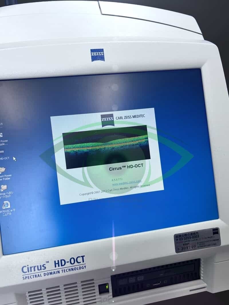
- Workstation / PC with Cirrus software
- Power cabling (as photographed)
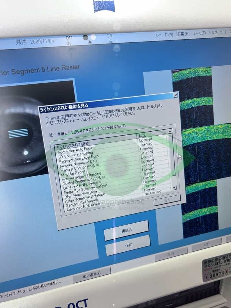
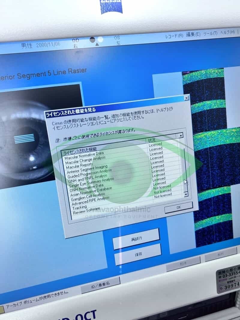
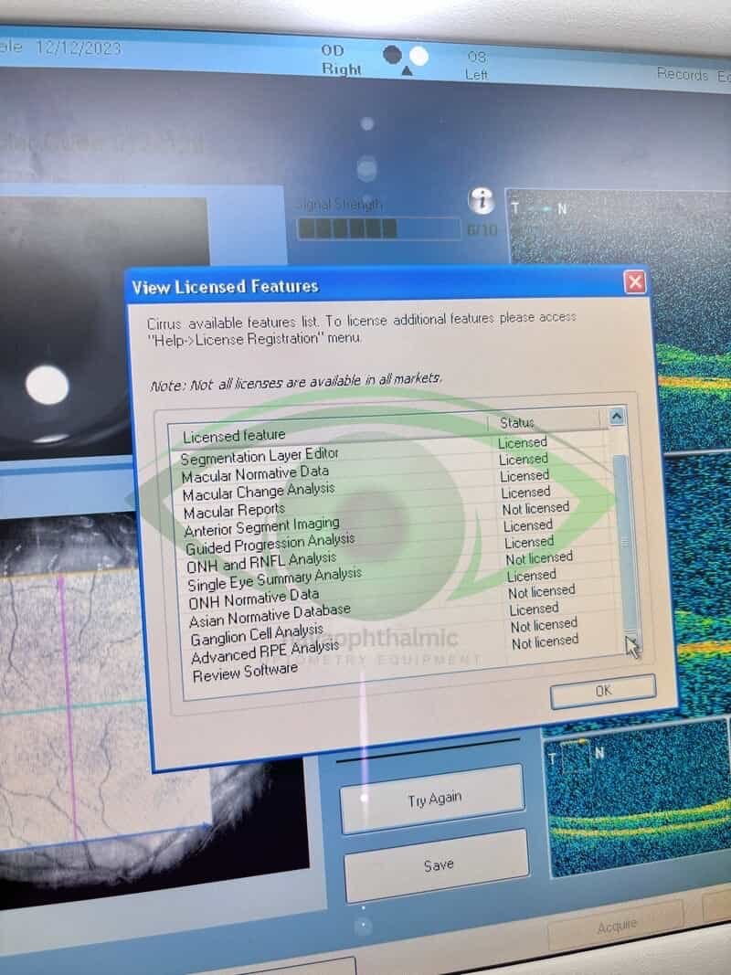
Licenses enable : Acquisition Auto Focus, 3D Volume Rendering, Segmentation Layer Editor, Macular Normative Data, Macular Change Analysis, Macular Reports, Guided Progression Analysis, ONH and RNFL Analysis, ONH Normative Data, Asian Normative Data, Ganglion Cell Analysis
Brochure:https://www.zeiss.com/content/dam/Meditec/us/download/certified-pre-owned-systems/Cirrus400Brochure__NewSoftwarePackage_CIR.3607.pdf