Description
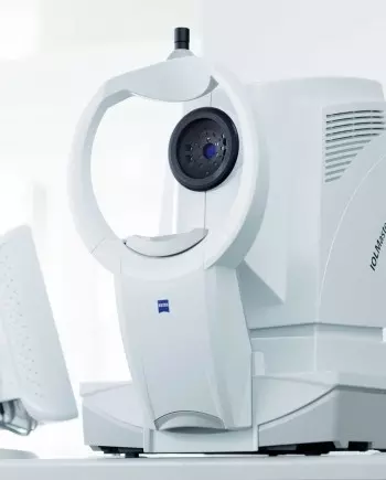
The CLARUS 700 from ZEISS is a complete ultra-widefield retinal camera. It helps eye care specialists take ultra-widefield images in true color. The camera offers excellent image quality and includes many features, such as fluorescein angiography.
The ZEISS CLARUS has transformed my ophthalmology practice. Its ultra-widefield imaging technology gives a clear panoramic view of the retina in true color. This reveals eye conditions that traditional methods often miss.
The clear and detailed images of the peripheral retina are very helpful. They let me find serious retinal diseases early. With the ZEISS CLARUS, I can diagnose and track these conditions with great confidence. This helps ensure my patients get the best care possible.
Dr. Aditya Kelkar – National Institute of Ophthalmology, Pune
Ultra-widefield fluorescein angiography serves as a highly valuable examination tool for assessing nonperfused retinal areas. The ZEISS CLARUS 700 HD is a special camera for eye imaging. It captures True Color, high-resolution images of the retina. It captures 133°1 in a single image and up to 267° with multiple captures, offering detailed views of the retina.
Equipped with both fluorescein angiography and live infrared imaging capabilities, the CLARUS 700 aids in the diagnosis and monitoring of retinal diseases. The fundus imaging camera features new technological advancements.
It includes PrecisionFocus, which helps see details quickly. QuickCompare allows doctors to compare changes in past patient visits. AutoBright lets ophthalmologists spend more time analyzing images and less time adjusting them.
Features
- HD fluorescein angiography
- True-color imaging
- Exceptional 7-micron resolution
- Advanced imaging modalities
- AutoBright (automatic brightness adjustment)
- GazePoint (automated fixation point)
- PrecisionFocus
- Integration with ZEISS Integrated Diagnostic Imaging
The ZEISS CLARUS 700 is a single UWF platform. It addresses many challenges that clinicians encounter in their clinics. True color, FAF blue, FAF green, and FA are all available on the CLARUS 700. It also offers external photography and infrared imaging (Figure 1).
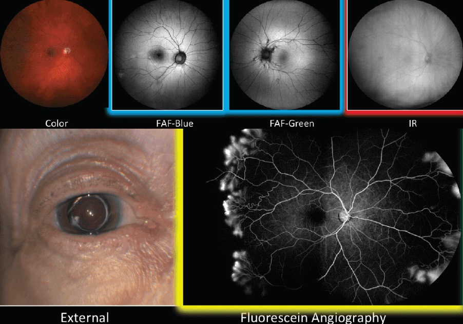 Figure 1. Multiple modalities are available on the single platform of the ZEISS CLARUS 700.
Instead of using a single white light for UWF true-color fundus photos, the CLARUS 700 utilizes three colored LEDs. These LEDs help scan the retina during color fundus photography. The platform uses a red LED (585-840 nm), a green LED (500-585 nm), and a blue LED (435-500 nm). This setup produces a fundus photograph that appears natural, much like what you would see with direct observation.
The algorithmic adjustment removes lash and lid artifacts, providing a cleaner image for assessment. The accuracy and resolution are like those of a traditional 45° camera. However, it offers a widefield view of up to 200° with montaging.
The CLARUS 700 has three features that make it easier to use. These features also increase the data available in the clinic.
PrecisionFocus technology enhances our ability to examine a specific region of interest without compromising resolution in other areas. AutoBright software optimizes an entire series of images (rather than optimizing each image individually) for brightness. GazePoint artificial intelligence (AI) technology facilitates montaging by identifying and using the optic nerve as a focal point, providing a superior UWF image.
PrecisionFocus
PrecisionFocus is particularly useful in eyes that are not emmetropic. In these eyes, features close to the center are clear. Images on the edges are seen in lower detail. With PrecisionFocus technology, details in the foveal center and the periphery are both rendered in high resolution (Figure 2).
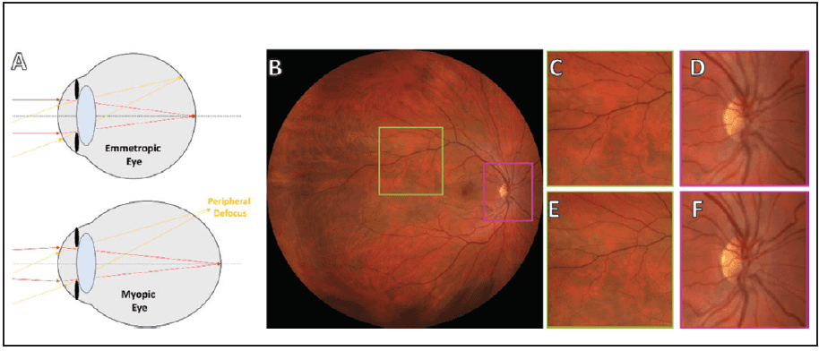 Figure 2. Peripheral defocus is a common issue encountered when imaging non-emmetropic eyes (A). Looking at the vessels in a 90° image of a myopic eye can make the images less sharp. This effect is noticeable near the foveal center.
Without PrecisionFocus technology, the vessels (C) are in a higher resolution than the area near the optic nerve (D). When PrecisionFocus technology is used, sharpness is maintained peripherally (E) and centrally (F).
AutoBright
Images that are too dark lack sufficient contrast (Figure 3, top row). Images that are too bright lose details (Figure 3, bottom row). AutoBright technology on the CLARUS 700 changes images during processing. This ensures that images have good contrast and detail, making them useful for the clinician.
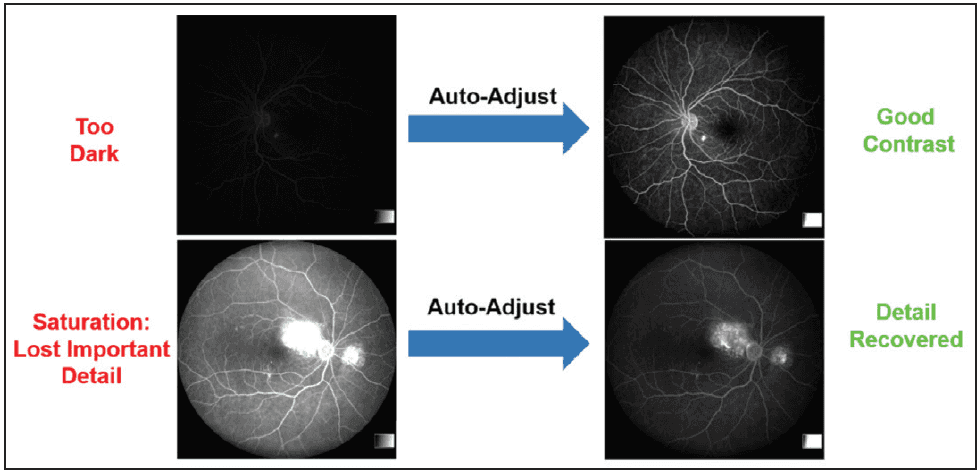 Figure 3 shows how AutoBright technology on the CLARUS 700 improves images. It makes dark images (top row) brighter and reduces saturation in overly bright images (bottom row). This helps ensure the imaging report is easy to read.
GazePoint
GazePoint technology on the CLARUS 700 leverages the power of AI to create fully detailed montage UWF images. When patient fixation is imperfect, the resulting image may be suboptimal. With GazePoint, the optic nerve becomes the point of fixation, resulting in a sharper image (Figure 4).
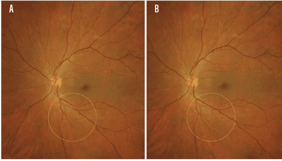 Figure 4. UWF images that are not perfectly fixed can show results like a break in a vessel (A, yellow circle). After applying GazePoint, artificial intelligence adjusts the image, resulting in a readout that shows vessel continuation (B, yellow circle).
CASE EXAMPLE
In this patient with severe nonproliferative diabetic retinopathy, limited details are available on standard 7-field imaging (Figure 5A). Using UWF imaging on FA helps us better understand the disease’s extent and the severity of leakage (Figure 5B). With help from PrecisionFocus, AutoBright, and GazePoint technologies, the CLARUS 700 produces a clear image. The image also has good contrast.
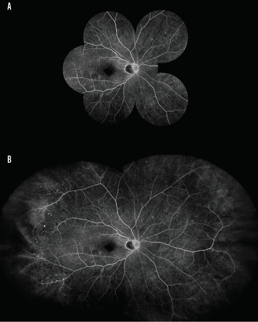 Figure 5. FA imaging of a patient with severe nonproliferative diabetic retinopathy reveals limited data when employing standard 7-field imaging (A). When UWF imaging is employed, the nature and extent of the disease is easily observed (B).
DOING MORE WITH LESS
In a busy clinic, efficiency and efficacy are key. The CLARUS 700 combines various types of tests on a single platform. This helps my clinic work efficiently. My patients can have several tests done quickly.
Software updates that provide clearer, more detailed images enable me to analyze my patients more effectively. They also let me customize treatments to meet each patient’s needs.
Technical Details
- Field of view: Wide field (133°), Ultra wide field (200°), Mounting (up to 267°)
- Optical resolution: 7.3 µm
- Minimum pupil diameter: 2.5 mm
- Working distance: 25 mm (from patient’s eye to front lens)
- Ametropia compensation: continuous -24 D to +20 D
- Weight: 23.6 kg
- Dimensions (W × D × H): 36.2 cm × 54.6 cm × 67.6 cm
Reference