Inhoudsopgave
Eye health is critical to overall well-being. However, people often overlook it until a problem arises. Regular eye exams are essential for early detection and prevention of severe eye conditions.
With modern advances, retinal images have revolutionized how eye care professionals diagnose and treat eye diseases. This cutting-edge retinal imaging technology offers unparalleled precision and insight, making it a cornerstone of advanced ophthalmic care.
Imagine diagnosing eye conditions before symptoms appear, preventing vision loss, and improving patient outcomes. Diagnostic retinal scans are making this possible.
Keep reading to explore how these advancements are reshaping eye health.
What Is a Digital Retinal Image?
A digital retinal image is a clear photo or scan of the retina. The retina is the light-sensitive tissue at the back of the eye. This intricate tissue converts light into neural signals, allowing the brain to interpret visual information.
Retinal imaging technology captures fine details of the retina, such as:
- Blood vessels
- The macula
- The optic nerve head
These insights are crucial for assessing overall eye health and diagnosing potential issues. The ability to visualize and analyze the retina with such precision has made this technology indispensable in modern ophthalmology.

How Does Retinal Imaging Work?
Understanding how retinal imaging operates is essential for both patients and practitioners. The process highlights the synergy between advanced technology and patient care, demonstrating its value in modern ophthalmology.
Equipment
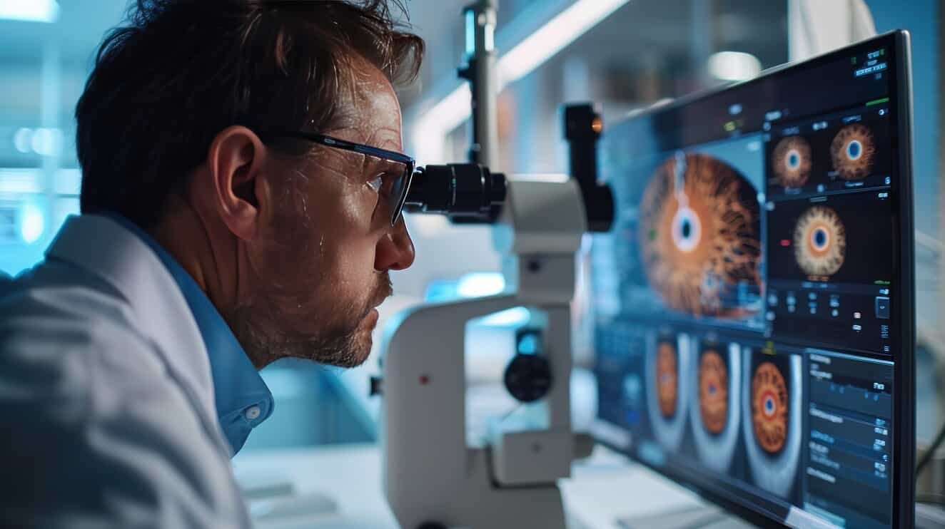
Retinal imaging relies on cutting-edge devices such as OCT (Optical Coherence Tomography) and fundus cameras. OCT uses light waves to create detailed cross-sectional images of the retina. It captures layers of tissue with micrometer precision.
This allows for in-depth analyzing of the macula and optic nerve head.
Fundus cameras offer wide views of the retina. They provide high-resolution images, ensuring that small problems in large areas do not get missed. Advanced models, like those offered by Nava Ophthalmic, integrate:
- High-resolution imaging
- Multi-modal capabilities
- AI-driven analysis
Process
Before diving into the procedure, it is crucial to understand why it matters. The process provides a seamless experience for patients while offering clinicians comprehensive data. Let’s look at the process step-by-step:
- Patients are guided to sit comfortably with their chin resting on a support to ensure stability
- They focus on a designated point, allowing the device to align and capture images
- A non-invasive beam of light scans the retina in seconds, producing clear and accurate visuals
- These images are processed and immediately available for analysis by the ophthalmologist or integrated software
The seamless procedure is painless and fast.
Voordelen van digitale netvliesfotografie
As technology evolves, new advancements are setting benchmarks in eye care diagnostics. Modern ophthalmic imaging trends highlight numerous benefits.
Vroege Opsporing van Oogziekten
Modern imaging tools provide the precision and clarity that allow for early detection of conditions like diabetic retinopathy, glaucoma, and macular degeneration. These conditions often develop silently, progressing without noticeable symptoms until significant damage has occurred.
Early detection through retinal imaging technology allows ophthalmologists to identify even subtle changes in the retina. This provides a clear window into the progression of these diseases. An active way to protect vision and keep eyes healthy exists. This approach allows for treatment before severe damage happens.
Early intervention reduces the risk of permanent vision loss by allowing timely treatment, such as:
- Lasertherapie
- Injections
- Surgical procedures
These treatments depend on the condition and its severity.
Better Diagnostic Accuracy
High-resolution retinal images reveal even minute abnormalities, such as tiny hemorrhages, microaneurysms, or subtle changes in retinal thickness. These intricate details are critical for diagnosing complex conditions early and accurately.
For instance, early signs of diabetic retinopathy can appear as microaneurysms or small leaks in the retinal blood vessels. On the other hand, glaucoma may show a subtle thinning of the optic nerve fibers.
Identifying these markers early on allows for timely interventions, which can:
- Reduce the risk of vision loss
- Improve long-term outcomes
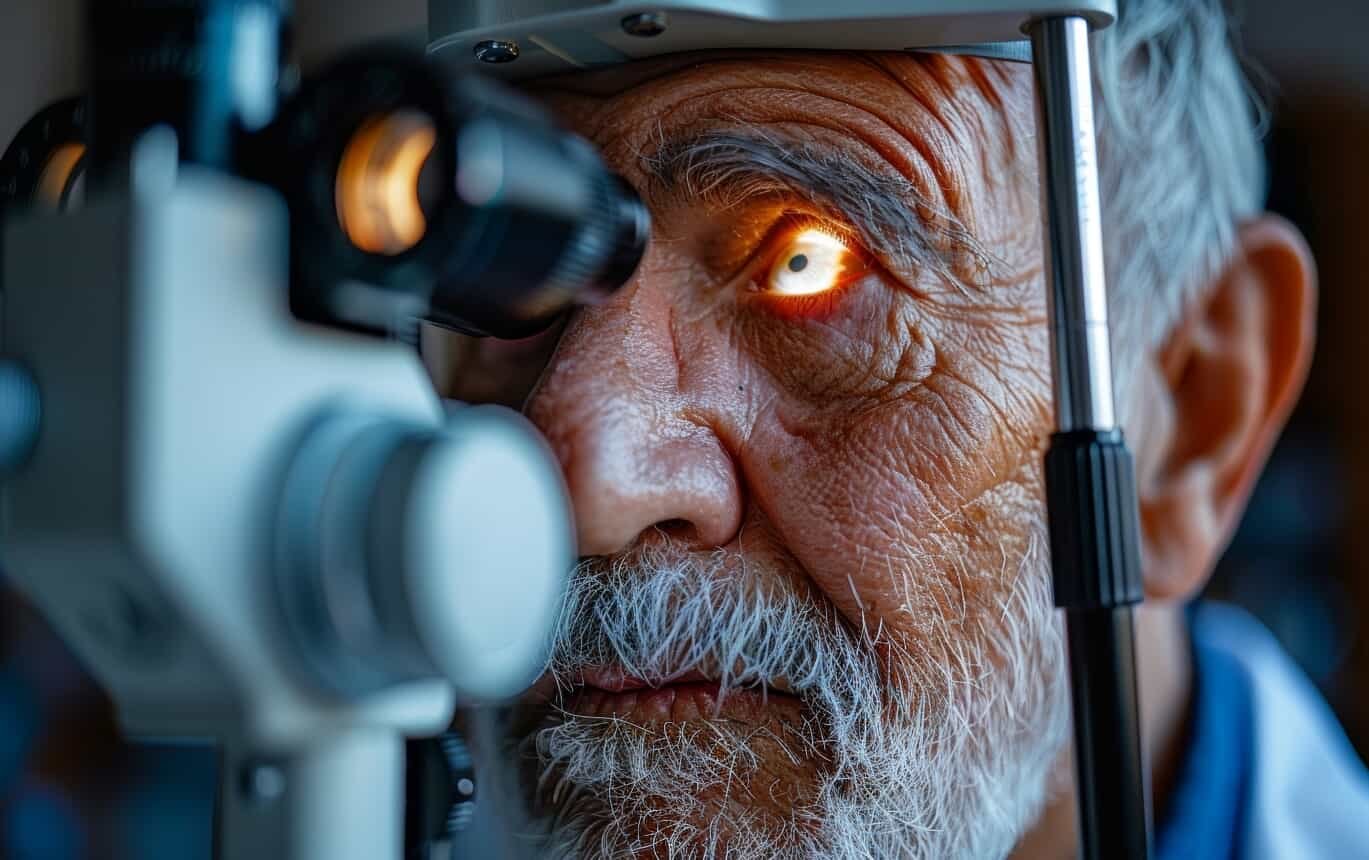
Eye doctors can give clear explanations to patients. They show visual evidence of the patient’s condition. This includes detailed images that highlight the affected areas. This approach:
- Fosters better understanding
- Builds trust
- Empower patients to participate in their care plans.
Comprehensive Records
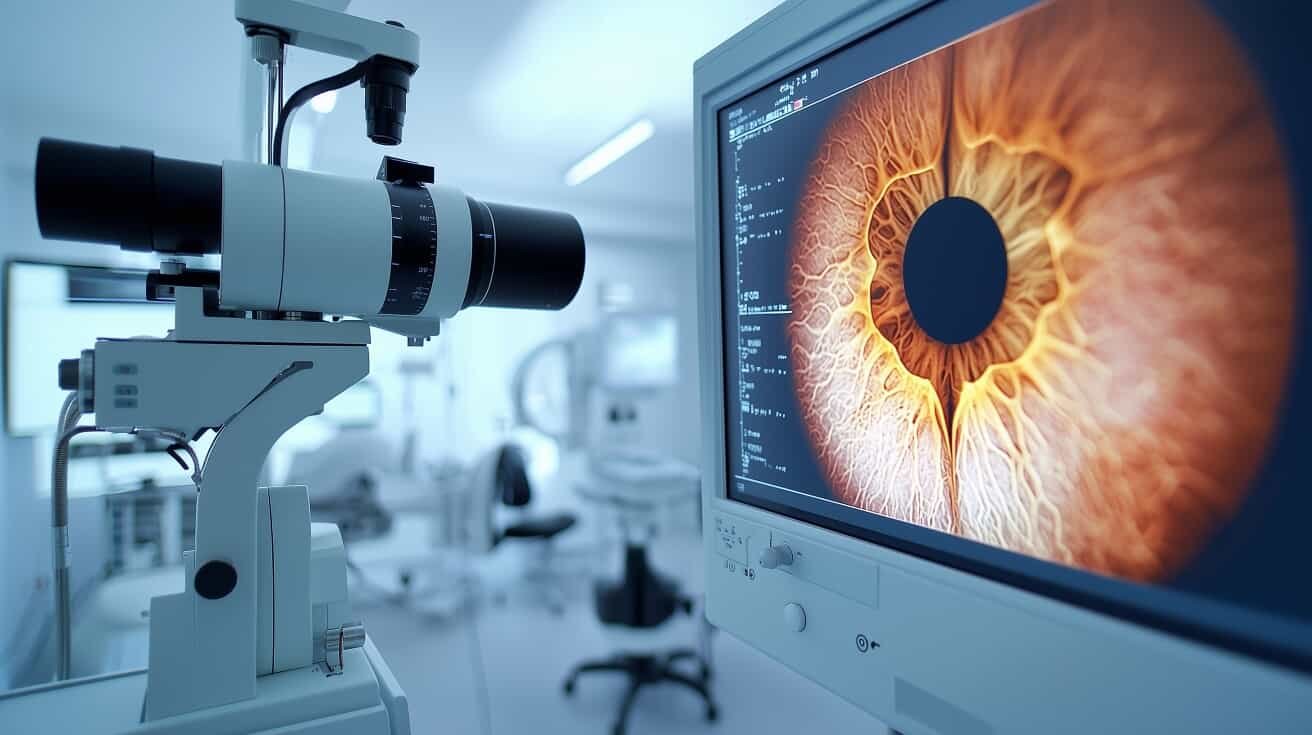
Images are stored digitally for comparison over time. This creates a comprehensive database for each patient’s eye health. A process allows for historical tracking and helps establish baselines highlighting subtle deviations.
Ophthalmologists can use these insights to watch for slow changes in the retina. This includes spotting microvascular problems in diabetic retinopathy or early damage to the optic nerve in glaucoma. The ability to compare past and present images ensures a dynamic understanding of a patient’s condition.
You can track the progression of conditions with precision. It also enables timely interventions, such as:
- Changes to medication
- Surgical planning
- Preventive measures tailored to the patient’s specific needs
This ensures a proactive and personalized approach to eye care.
Non-Invasive Procedure
Unlike older methods, diagnostic retinal scans are painless and often require no dilation. This makes the procedure more comfortable for patients. It eliminates the discomfort and light sensitivity usually associated with traditional dilation.
Reducing patient anxiety is critical, especially for those who may already feel apprehensive about medical procedures. The shorter recovery time allows patients to resume daily activities immediately after the scan.
Non-invasive scans help high-risk patients and those with specific medical conditions. They can safely undergo the procedure. This mix of comfort, convenience, and safety makes retinal scans a great choice for regular eye exams. They are also good for keeping track of long-term health issues.
Patient Education
Visual aids from scans help patients see their conditions. They provide a clear view of what is happening inside their eyes. Ophthalmologists can explain complex situations in simple, visual terms by showing patients high-resolution retina images.
For instance, a patient with diabetic retinopathy can see how damaged blood vessels impact their vision. This approach enhances understanding and encourages patients to participate actively in their treatment plans. The result is, ultimately, improved compliance and long-term outcomes.
Streamlined Workflow
Clinics equipped with advanced retinal imaging technology experience improved efficiency by automating many aspects of the diagnostic process. These technologies reduce the time required for image acquisition and analysis, enabling faster patient turnaround and optimized scheduling.
Integrating electronic medical records (EMRs) ensures healthcare providers seamlessly update patient data. This allows for better coordination across teams and departments.
Cost Savings
Early detection prevents costly treatments for advanced diseases by identifying problems before progressing to critical stages. For example, detecting diabetic retinopathy early on may avoid the need for expensive surgeries or vision-saving injections.
Early diagnosis of glaucoma can prevent irreversible damage that leads to significant medical expenses and reduced quality of life. By leveraging advanced retinal imaging technology, clinics can reduce the financial burden on patients and healthcare systems.
Disadvantages of Retinal Scanning
Retinal scanning technology offers numerous benefits, but specific challenges persist. Understanding these drawbacks helps clinics and patients make informed decisions about its use.
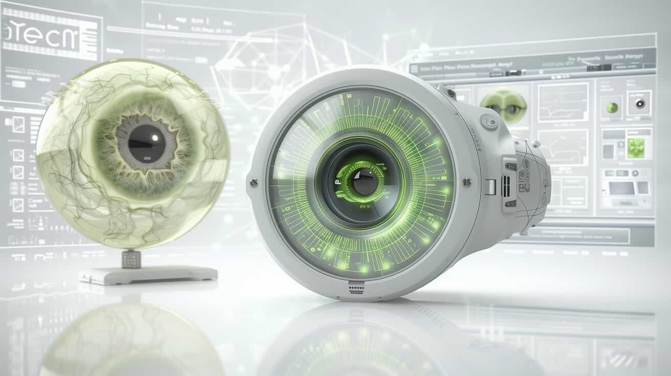
High Initial Investment
The cost can be higher for clinics and patients. For clinics, investing in advanced retinal imaging technology often requires a significant upfront expense, including:
- Purchasing equipment
- Integrating software
- Training staff to use the devices the correct way
These costs can also impact patients. Some clinics may need to charge higher fees to cover their operational expenses.
However, many clinics find that the long-term benefits outweigh the initial investment.
Accessibility Challenges in Underserved Areas
Access to retinal imaging technology can be challenging in underserved areas. Financial problems and a lack of specialized clinics make it hard for patients to receive care.
Limited advanced equipment is also available. This combination creates challenges for those seeking medical help. We must use portable devices, telemedicine, and funding programs to fix these gaps. This will help everyone get good eye care.
Why Retinal Imaging Is Important
Retinal imaging is a crucial tool for proactive eye care. Regular imaging helps track changes and detect potential issues before they worsen.
The technology helps not just individuals but also public health efforts. It finds patterns in eye health trends for the whole population.
How Often is Retinal Imaging Needed?
Doctors recommend retinal imaging every 1-2 years during routine eye exams for patients with no known issues. This frequency helps set a baseline for their retinal health and allows for detecting small changes that may show early signs of eye problems.
Those with conditions like diabetes, hypertension, or a family history of eye diseases may need more frequent scans. They are at a higher risk for developing conditions like:
- Diabetische retinopathie
- Hypertensive retinopathy
- Glaucoom
These conditions can progress rapidly without early intervention. Regular imaging allows for close monitoring, enabling timely detection and treatment to prevent serious complications.
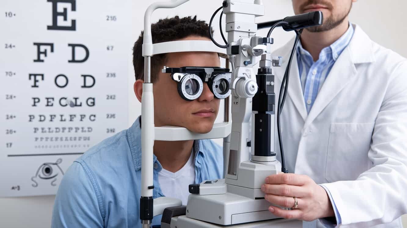
Common Eye Diseases Identified Through Retinal Imaging
Retinal imaging has become an indispensable tool for detecting various eye diseases. Each condition it identifies can have significant implications for vision and overall health, highlighting the importance of routine imaging.
Let’s dig a little deeper concerning the most common eye diseases.
Diabetische retinopathie
High blood sugar damages retinal blood vessels, leading to vision loss. This condition is often asymptomatic in its early stages, making routine imaging crucial for early detection and management. Retinal imaging reveals:
- Microaneurysms
- Hemorrhages
- Fluid accumulation
Glaucoom
Increased eye pressure damages the optic nerve, leading to gradual vision loss. Advanced imaging tools like OCT can detect early thinning of the optic nerve fibers. This happens before symptoms appear, allowing for earlier treatment.
Age-Related Macular Degeneration (AMD)
AMD affects central vision and is a leading cause of blindness in older adults. Imaging helps distinguish between dry and wet AMD, guiding treatment plans to slow the disease and protect vision.
Retinal Detachment
Early detection through imaging can prevent severe outcomes. Retinal imaging helps doctors act quickly to save or improve vision. It finds small tears or detachments before they get worse.
Eye Health Advancements and Trends
The latest eye health advancements revolutionize diagnostic and treatment capabilities in ophthalmology. By incorporating cutting-edge technologies, these advancements enhance:
- Accessibility
- Precision
- Patient care
Portable Devices
Tools like the Volk Pictor allow imaging in remote locations. These portable devices expand access to quality eye care in underserved regions by overcoming geographical and logistical barriers. Their compact design and ease of use make them invaluable for:
- Mobile clinics
- Community outreach programs
- Rural healthcare settings
These devices provide precise and reliable images. They help find serious health issues in patients who may not get this care otherwise.
Advanced Equipment
Nava Ophthalmic supplies state-of-the-art devices, such as the Zeiss Clarus 500, for precise diagnostics. These devices provide unparalleled accuracy and enable clinicians to capture detailed retina images for early diagnosis and treatment.
De RetCam 3, for instance, is ideal for pediatric retinal imaging. It offers specialized solutions for unique needs.
Its ability to safely take images of the delicate eyes of infants and young children makes it a key tool. The RetCam 3 helps detect conditions like retinopathy of prematurity and other eye disorders in children. Its advanced imaging capabilities allow doctors to identify and treat even the most subtle abnormalities.
Telemedicine Integration
Retinal images can be shared with specialists for remote consultations, facilitating collaboration between general ophthalmologists and subspecialists. This capability enhances diagnostic accuracy and ensures that patients in rural or underserved regions receive high-quality care.
The Role of Patient Engagement in Retinal Imaging
Advanced imaging tools are now designed with patient experience in mind. These tools provide high-quality visuals that simplify complex medical information, making it easier for patients to comprehend their eye health status.
Patients can better understand their conditions by showing areas of concern or improvement. This helps them see why following treatment plans is essential. Educated patients are more likely to:
- Follow recommended therapies
- Attend regular follow-ups
- Actively participate in their care
Unlock the Power of Retinal Images for Better Eye Care
Incorporating modern retinal images into your practice enhances diagnostic accuracy and improves patient care.
At Nava Ophthalmic, we focus on providing advanced equipment. We offer tools like the Cirrus HD-OCT 5000 and RetCam 3. These help ophthalmologists give the best care possible.
We offer a comprehensive range of reliable, advanced ophthalmic solutions tailored to meet the specific needs of clinics. This ensures superior diagnostic precision and exceptional patient outcomes.
Browse our digital retinal imaging products today and discover how they can transform your practice.

Matthew Strachovsky, M.D.
Dr. Strachovsky's undergraduate opleiding begon in Boston, Massachusetts aan de Boston University en werd voltooid aan de Stony Brook University in Long Island, NY. Daar is hij Summa Cum Laude afgestudeerd, met een Bachelor of Science diploma in Biologie en speciale erkenning voor academische prestaties.
Hij vervolgde zijn opleiding aan de Stony Brook School of Medicine en studeerde af met de extra aanduiding van het "MD met erkenning" programma. Hij werkte als stagiair interne geneeskunde in het Winthrop University Hospital in NY en volgde een specialisatie oogheelkunde aan het Stony Brook University Hospital, waar hij in zijn laatste jaar optrad als hoofd-assistent (Chief Resident). Hij voltooide zijn fellowship training in vitreoretinale ziekten met een belangrijke focus op de diagnose en behandeling van retinale vaatziekten onder leiding van Dr. Michael O'Brien aan Koch Eye Associates in Rhode Island.
Dr. Strachovsky heeft onderzoek gepresenteerd op de jaarlijkse bijeenkomst van de Association for Vision and Research in Ophthalmology en artikelen gepubliceerd in tijdschriften zoals Investigative Ophthalmology and Visual Science en The Journal of Neuro-ophthalmology.
De professionele interesses van Dr. Strachovsky omvatten het beheer van leeftijdsgebonden maculadegeneratie en diabetische oogziekten. Hij is Board Certified in Oogheelkunde en lid van de American Academy of Ophthalmology, American Society of Retina Specialists, Young Ophthalmologist Network en Leading Physicians of the World.
"Ik geloof dat de arts-patiëntrelatie belangrijker is dan ooit. Als oogarts kan ik patiënten helpen en een basis van vertrouwen, kennis en professionaliteit opbouwen als het gaat om oogzorg.".
 English
 Spanish
 German
 Dutch
Dutch French
 Portuguese