Table of Contents
Digital retinal imaging is a device that helps in the early detection of eye conditions. Early screening can save you from problems and reverse situations such as glaucoma. The method involves the use of digital cameras to view and manipulate images.
Do you go for eye screening as much as you should? We often do not see the need to take eye exams regularly. Even if your eyes seem to be healthy, you should not overlook these tests. Most eye diseases develop gradually for years without revealing any symptoms. Optometrists reveal that most people who get blind later in life develop it out of later detection of eye diseases.
What is digital retinal imaging?
Doctors perform a comprehensive eye examination to conduct an in-depth analysis of your eyes. They do this with the help of digital cameras, which provide a good view of the eyes. Digital imaging is, therefore, the process of capturing digital images of the inner parts of the eye. The retinal camera can focus on the optic nerve, retina, blood vessels as well as macula.
You can screen a patient for any eye disease and compare them for future analysis with these images. If the pictures show eye diseases, you should take another higher quality test to confirm your suspicions.
Digital retinal imaging goes beyond the routine checkup in that you can view the whole retina from different angles to quickly detect a disease. The instrument can give you a clear picture of the retinal periphery.
Modern forms of imaging make doctors comprehend eye conditions such as macular degeneration and diabetic retinopathy better than in the past. Emerging techniques are used in screening, assessing, and monitoring a patient’s eyes to facilitate treatment more efficiently.
Our discussion aims at helping doctors find ideal digital retinal imaging equipment. We will also help patients understand how they can monitor their progress by using instruments such as the best tonometer for home use. It takes the efforts of both parties to treat eye conditions and reduce the risk of diseases. Find out more!
Detecting early eye diseases
Experts report that the number of patients losing eyesight from illnesses is increasing. Age is a factor that causes some eye diseases. This means that every person beyond 40 years is at risk of developing eye diseases like cataracts or macular degeneration. Early detection can help the doctor catch any eye problems before it advances.
Start by making yearly visits to the eye clinic as you age and make them more frequently if you are experiencing any changes in your eyes. Apart from screening, it would help if you also made some lifestyle changes such as monitoring your diet and quitting smoking to improve the condition of your eyes. Get the best tonometer for home use to keep track of your eye pressure without the presence of your doctor.
Advantages and Disadvantages Digital Retinal Imaging

Eye doctors have a wide range of imaging instruments to select from. Some of them are sophisticated and need special skills to use, while others are simple to manipulate. While choosing this equipment, you should weigh its merits against limitations to select an ideal type.
Imaging tools eliminate the need for pupil dilation to view the inner parts of the eye. You may not need to use eye drops on the patient to see the parts. This means that the instruments do not need any preparation. They make the process painless and comfortable for the patient. They are safe to use as they utilize infra-red light. You can also conduct the test fast and still acquire high image resolution.
Using these tools gives you a clear picture of the patient’s eye health and makes it easy to compare notes with other physicians to develop an accurate report. They help you view the retina from a wide angle to detect diseases early enough. Most of the devices come with multiple filters that you can use to enhance images and see fine details of the different eye parts.
Though digital retinal imaging is a blessing for eye doctors, it has some limitations. It is hard to detect a disease in a patient if the retina is bleeding. The tools also come at a high cost, meaning you have to compare prices before purchasing one. Such devices also come with specific challenges, which may include color inaccuracy and insufficient image acquisition.
Here are the top 10 Digital Retinal Imaging Products on the market.
1. Zeiss Clarus 500 ($20,000.00) – a User-friendly imaging tool
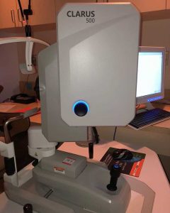
ZEISS Clarus 500 satisfying patient encounter provides images free of obstructions, such as lids and lashes, and demands fewer recaptures for a thorough eye exam. Many eye practitioners select this instrument due to how easy it is to use without special skills.
ZEISS Clarus 500 helps in the management of eye diseases as it provides high-resolution images of the retina. Zeiss Clarus 500 reduces some of eye doctors’ challenges since it delivers accurate images for proper analysis.
Features
- 7-micron resolution
- Auto—merge
- LEDs
- Chinrest
The micron resolution allows you to view areas such as the retinal periphery to acquire quality images. This can help you detect both peripheral and macular diseases. Thanks to the auto-merge feature, you can capture true-color pictures from different angles. It also utilizes LEDs to help you capture images in their real form.
Check our Review about ZEISS Ophthalmic Equipment
The manufacturer of this digital camera has both the doctor and the patient in mind. That is why they incorporate a sleek design to ensure that the patient remains comfortable during the eye test. They equip it with a chin rest to ensure that the patient remains still and relaxed while acquiring images.
You also get a live infrared preview to optimize alignment. This ensures that you can capture images without interference from the patient’s lashes and lids. The patient is also not exposed to excessive flashes as you capture images.
2. Optos California ($25,000.00) – A compact tabletop imaging device
Some patients have small pupils making it hard for an eye doctor to view the inner parts. Optos California solves such problems since it conducts imaging through small pupils and cataracts.
Optos California facilitates the capture of ICG and angiography images eliminating manual switching of modalities.
Features
- Angiography modes
- Proview software
- ICG imaging modality
- Cloud-based archiving
The users of this digital camera reveal that it can capture retinal images in different modalities such as fundus photography. It also comes with a small footprint and a compact design that increases the ease of access to patients. The inclusion of ergonomics makes it comfortable for patients to rest.
The producers equip this imaging tool with the Proview software, which allows it to display wide-field scans more consistently than other devices. This increases accuracy since it can show the anatomic features of a patient’s retina. The proprietary optical hardware enables you to acquire high-resolution images.
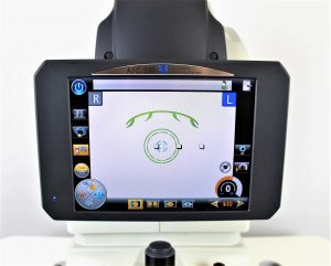
3. Centervue Eidon ($11,450.00) – Versatile and user-friendly device
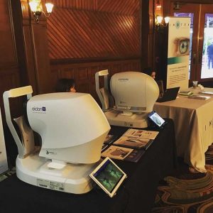
Are you looking for actual color images from a digital retinal imaging device? Centervue Eidon is a suitable choice. It utilizes white light illumination with confocal imaging to generate color images.
Centervue Eidon merges color channels to give you orange retinal photos. Eye doctors say that white light illumination provides precise details of the retina and optic nerve.
Features
- Confocal image options
- Manual and automated modes
- Compact design
- Joystick
The device provides high-resolution images due to the inclusion of different options like red-free and infrared. It can image through media opacities and cataracts and detect problems in a patient’s eyes.
You can run this DRI tool using either the manual or automated modes. It can cover up to 110 degrees while using the automatic mode and 150 degrees in the manual mode. The joystick allows you to adjust the equipment from automatic to manual mode, thus facilitating a customized focus.
You don’t need advanced skills to use them during eye exams. It aligns well with the pupil of your patient so that you can focus on the retina without any hindrances. It comes in a small print design to increase comfort for the patient.
Using this instrument helps you view the retinal image in its precise form. The multiple imaging modalities make it versatile, and the software interface is easy to understand.
4. Volk Pictor Plus ($6,666.00) – A mobile imaging tool
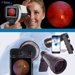
Pictor Plus is a fundus camera that is used to screen patients for conditions such as diabetic retinopathy.
Pictor Plus is a compact design that is lightweight to increase portability. Most eye doctors who go for missions in different regions utilize this device since it is not fixed.
Features
- Field of view: 40 degrees
- Two modules
- Eyecap
- Weight:1b
The digital retinal imaging equipment features two modules to help you view the eye’s external structure and the retina. It comes with an eye cap which makes the camera stable for you to capture images quickly. It also improves the patient’s experience by minimizing stray light.
Apart from being portable, the fundus camera generates high-resolution photos. It offers a 40-degree field of view. You can upload the images you capture to your computer since they are compatible with software programs such as EMR solutions. It does not limit you to work during the day since you can still capture images at night.
5. Retcam 3 ($23,999.00) -it is safe and fast
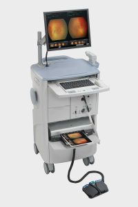
A digital retinal camera should give you stunning images to help you analyze the eye condition of your patients. Retcam 3 enables you to achieve this within minimal time. You can capture full-color pictures and use them to assess both the anterior chamber and retina.
Features
- Console design
- Keyboard
- Mouse
- Interchangeable lenses
It comes in the form of an ergonomic handpiece to enhance manipulation and make it easy for you to capture images. The weight increases portability and helps you conduct eye exams anywhere. Most users love the design of Retcam 3, which features an extended work surface and a keyboard and mouse for ease of use.
Feel free to interchange the lenses to acquire intricate details from your patient’s eye. The longitudinal thumbnail allows you to display the captured images for analysis. You can also view them using the full-screen mode available.
6. Centervue DRS ($7,999.00) – it is comfortable and user-friendly
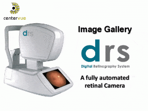
Do you deal with a large population in need of eye screening? Get this Centervue DRS that gives you high performance while reducing any complicated steps. It allows you to capture retinal images at a high resolution without sacrificing the comfort of your patients.
Features
- Telemedicine system
- Low power flash
- 7 LEDs
- Chinrest
Centervue DRS does not need a lot of training for you to learn how to use it. The telemedicine system ensures that you can manipulate images through the use of remote control. It utilizes a low-power flash to make patients as comfortable as possible.
The device is versatile in that you can use it for both diagnosis and screening. Taking retinal images using this device takes less than a minute, thanks to the touch screen mechanism. The manufacturers equip it with various LEDs to increase the quality of images you capture.
You can view the images using a tablet or PC as long as you have an internet connection. Upon detecting eye diseases such as glaucoma, you can recommend the best tonometer for home use to help patients monitor any eye pressure changes from home.
7. Optos Daytona ($19,550.00) – enables autofluorescence imaging

Capture ultra-wide images of the retina of your patient’s retina using the Optos Daytona. It is a digital instrument that utilizes a unique technology to increase efficiency. Though it is small, it can deliver high-resolution images within seconds.
Features
- Optus advanced software
- Chinrest
- LEDs
Optos Daytona contains autofluorescence capabilities, and it can view the retina from different angles. Unlike other cameras that only view the posterior pole, Optos can also display the peripheral retina. With this equipment, you can diagnose various eye conditions.
Using this machine, you can also detect any abnormalities in the peripheral area, such as holes, retinal tears, or detachments. It improves the patient’s experience since it is fast to test. You can later review the results with the patient as you educate them about their eye health.
8. Nidek AFC 330 ($5,040.00) – Low flash intensity tool
It is not easy to capture retinal images from young ones. It would help if you had a machine that keeps them still for a short time to obtain the photos. Nidek AFC 330 is ideal for patients, including children, since it is designed for special conditions. You can even use it on patients that have small pupils.
Features
- An in-built camera and computer
- Tilting screen
- Wi-Fi connection
This enables you to conduct high-resolution imaging as it positions itself automatically to capture images. It means that you don’t need a lot of expertise to operate the digital retinal imaging equipment. It is used in the screening of conditions such as diabetic retinopathy.
You can capture images without subjecting your patient to an intense flashlight. The process is both fast and silent. The tilting screen allows you to enter data fast and analyze your patient’s condition.
9. Canon CR-2 AF ($5,678.00) – The automatic digital camera
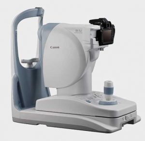
The Canon CR2 AF opacity suppression is what makes this digital camera unique from the rest. It is made using advanced technology to boost accuracy when it comes to retinal examinations. Feel free to view the images from the quick preview after capturing them.
Features
- Automatic and manual mode
- Auto capture
- Image error detection feature
- Digital cobalt mode
The software used eliminates any errors during focusing and confirms proper alignment to help you get accurate images. They come in a high resolution of about 20 megapixels. You also get wide-angle views to allow you to acquire contrast and details to diagnose any ocular diseases quickly. It enhances contrast by distinguishing the structure of the blood vessels from the surroundings.
Make use of the joystick to switch the automatic mode to the manual mode. The auto-capture feature makes it easy to capture images as soon as the eye is correctly focused. The auto-exposure feature regulates the intensity of infra-red light.
10. Topcon TRC 50 DX ($11,999.00) – delivers colored images

Get a chance to customize your settings to view retinal pictures from different angles using the Topcon TRC 50DX. It also allows you to adjust different filters as per the various procedures.
Features
- Control panel
- Aperture function
- Backlit panel
- Small pupil mode
- Color fundus
Topcon is a sophisticated fundus camera that improves the quality of retinal images to help diagnose ocular diseases. It incorporates advanced functions that make it versatile and easy to use. The aperture function is small to boost the focusing and encourage sharpness.
The machine supports different devices, including CCD and film cameras. Its control panel comes with a touchscreen that is easy to operate. It provides different flash intensity levels to make every patient comfortable. You can also use it in a dark area, thanks to the backlit panel available.
Conclusion
The use of digital retinal imaging makes it possible for optometrists to detect ocular diseases early enough to prevent people from going blind. These machines increase accuracy when it comes to diagnosing diseases such as glaucoma, macular degeneration, and diabetic retinopathy.
You can easily detect any fluctuations in the retina of your patient and store electronic records for future assessment. We should all pay attention to eye care and undergo examinations regularly. Ensure you get the best tonometer for home use to reduce the visits to the clinic.
Every eye doctor in the field should check out the models above and choose the ideal type. It will help you save time and get clear images for proper analysis.

Matthew Strachovsky, M.D.
Dr. Strachovsky's undergraduate training began in Boston, Massachusetts at Boston University and was completed at Stony Brook University in Long Island, NY. There he graduated Summa Cum Laude, obtaining a Bachelor of Science degree in Biology with special recognition for academic achievement.
He continued his education at Stony Brook School of Medicine and graduated with the additional designation of the "MD with Recognition" program. He worked as an intern in Internal Medicine at Winthrop University Hospital in NY and pursued a residency at Stony Brook University Hospital in Ophthalmology acting as Chief Resident in his final year. He completed his fellowship training in Vitreoretinal disease with a major emphasis on the diagnosis and management of retinal vascular diseases under the direction of Dr. Michael O'Brien at Koch Eye Associates in Rhode Island.
Dr. Strachovsky has presented research at the annual Association for Vision and Research in Ophthalmology meeting and published articles in journals including, Investigative Ophthalmology and Visual Science and The Journal of Neuro-ophthalmology.
Dr. Strachovsky's professional interests include the management of Age-Related Macular Degeneration and diabetic eye disease. He is Board Certified in Ophthalmology and a member of the American Academy of Ophthalmology, American Society of Retina Specialists, Young Ophthalmologist Network, and Leading Physicians of the World.
" I believe that the physician/patient relationship is more important than ever. Being an Ophthalmologist allows me to help patients and build a foundation of trust, knowledge, and professionalism when it comes to eye care".