Table of Contents
Vision loss and eye-related diseases are a growing concern globally. According to the World Health Organization, over 2.2 billion people have vision impairment, with at least 1 billion of these cases either preventable or yet to be addressed.
This alarming statistic indicates the critical need for regular eye check-ups and access to advanced diagnostic technologies like digital retinal imaging. Many eye conditions can be managed or treated with early detection, preserving millions of people’s vision worldwide.
Digital retinal imaging is among the most potent tools for preserving vision. This innovative technology is essential in identifying early signs of severe eye conditions, often before patients experience symptoms. In this comprehensive guide, we will explore digital retinal imaging, how it works, its benefits, and why it is a vital part of modern eye care.
As the demand for early detection of eye diseases grows, digital retinal imaging and optical coherence tomography (OCT) are becoming crucial in protecting and preserving vision. Read on to discover how this advanced technology can enhance your eye care routine and help you safeguard your vision.
What Is Digital Retinal Imaging?
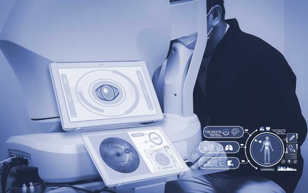
Digital retinal imaging is a non-invasive diagnostic tool that captures high-resolution images of the retina, optic nerve, and surrounding blood vessels. By examining these structures in detail, ophthalmologists can identify early signs of eye diseases like macular degeneration, glaucoma, and diabetic retinopathy.
Unlike traditional eye exams, which provide only a limited view of the eye, digital retinal imaging offers a comprehensive and detailed look at the retina’s various layers. This allows doctors to detect changes in the eye far earlier than with other methods, enabling timely treatment and potentially preventing permanent vision loss.
Optical Coherence Tomography (OCT)
 A related and equally important technology is optical coherence tomography (OCT). OCT provides cross-sectional images of the retina, enabling an even more detailed examination of the eye’s inner layers. This non-invasive eye scanning method allows doctors to detect subtle changes that may indicate the onset of diseases like macular degeneration or diabetic retinopathy.
Key elements of digital retinal imaging include:
- Non-invasive procedure: The imaging process does not require direct contact with the eye
- Advanced technology: High-resolution imaging provides detailed pictures of the retina
- Ongoing monitoring: Images can be stored for future reference, allowing doctors to track changes over time
Together, digital retinal imaging and OCT provide a powerful combination for diagnosing, monitoring, and managing eye diseases.
Benefits of Digital Retinal Imaging
The benefits of digital retinal imaging extend beyond simple eye exams, offering deeper insights into retinal health and early disease detection. Let’s take a closer look at some of the key advantages.
Early Detection of Eye Diseases
One of the most significant advantages of digital retinal imaging is its ability to detect eye diseases before symptoms arise. It is especially critical for conditions like glaucoma, which may not present noticeable symptoms until significant vision loss has already occurred.
Improved Patient Understanding and Engagement
One of the most powerful features of digital retinal imaging is its ability to enhance communication between doctors and patients. By showing patients detailed images of their retinas, doctors can explain the presence (or absence) of conditions in a way that is easy to understand. This helps patients feel more informed and involved in their eye care, leading to better adherence to treatment plans.
Additionally, digital images provide a valuable record that can be used for long-term monitoring. By comparing images from one year to the next, doctors can track the progress of conditions like diabetic retinopathy, macular degeneration, and glaucoma, making it easier to adjust treatment plans as needed.
Non-Invasive and Painless
For patients, one of the most appealing aspects of digital retinal imaging is that it is completely non-invasive and painless. Unlike older methods that require dilating the pupils or using uncomfortable devices to examine the eye, digital retinal imaging simply requires the patient to sit still. At the same time, the machine captures images of the eye.
Because it’s painless and quick, this imaging method encourages patients to return for regular check-ups, which is critical for maintaining long-term eye health.
How Does Digital Retinal Imaging Work?
 Digital retinal imaging is a straightforward process that usually takes a few minutes. Here’s how it works.
Preparing for the Scan
The patient is asked to sit comfortably in front of the imaging device. In most cases, there is no need for dilation, making the procedure more convenient.
Image Capture
The imaging machine uses advanced technology to capture high-resolution images of the retina and surrounding structures. The process is quick, usually taking less than a minute.
Reviewing the Images
Once the images are captured, the ophthalmologist reviews them for any signs of abnormalities. With OCT, cross-sectional images of the retina can also be examined to detect subtle changes that may indicate early disease.
Data Storage
The images are saved for future reference, allowing for comparison with subsequent scans. This makes it easier for doctors to track the progression of eye diseases over time.
How Often Should You Get Retinal Imaging?
The frequency of retinal imaging depends on your risk factors. For most people, an annual eye exam that includes digital retinal imaging is recommended. However, those with higher risk factors, such as diabetes, high blood pressure, or a family history of eye disease, may need to undergo imaging more frequently.
Regular imaging is essential because it allows doctors to detect changes in the eye before they lead to noticeable symptoms or vision loss. It’s important not to wait for symptoms to appear-regular imaging can make all the difference in protecting your vision.
Digital Retinal Imaging for Specific Conditions
Digital retinal imaging plays a critical role in managing several common eye conditions. Here are some examples of how this technology can be used to diagnose and monitor specific diseases.
Macular Degeneration
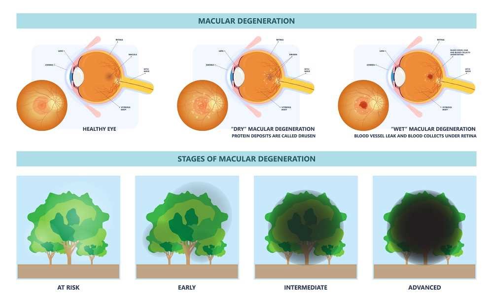
Macular degeneration, especially age-related macular degeneration (AMD), remains a primary cause of vision loss among individuals aged 50 and older. Digital retinal imaging plays a vital role in detecting early signs of this condition by capturing detailed images of the macula, the central part of the retina responsible for sharp, detailed vision. Doctors can implement strategies to slow disease progression and preserve vision when identified early.
Common interventions include:
- Lifestyle changes, such as adopting a healthy diet and reducing exposure to blue light
- Medications, including anti-VEGF injections, prevent abnormal blood vessel growth
- Other treatments, such as laser therapy or specialized eye supplements
By monitoring the progression of AMD through digital retinal imaging, doctors can adjust treatment as needed to maintain optimal eye health.
Glaucoma
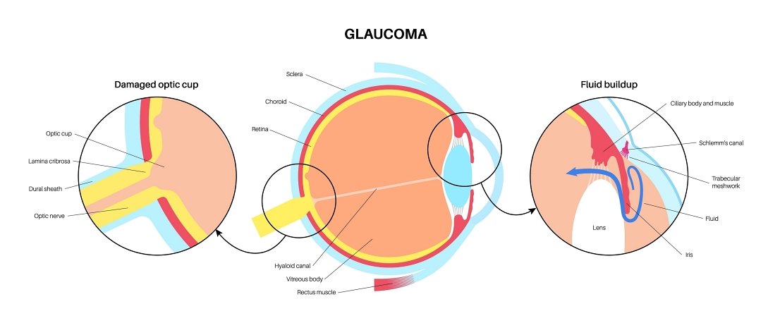
Glaucoma is often referred to as the “silent thief of sight” because it typically advances without apparent signs until substantial loss of vision happens. With digital retinal imaging, early detection is possible by capturing precise images of the optic nerve, which helps identify abnormalities before they cause irreversible damage.
Additionally, optical coherence tomography (OCT) offers enhanced diagnostic insights by providing detailed 3D images of the optic nerve’s layers. This capability allows doctors to detect thinning of the nerve fibers, a hallmark of early-stage glaucoma.
When caught early through non-invasive eye scanning, patients can benefit from timely interventions, including:
- Medicated eye drops
- Laser therapy
- Surgery
These interventions prevent further deterioration.
Diabetic Retinopathy
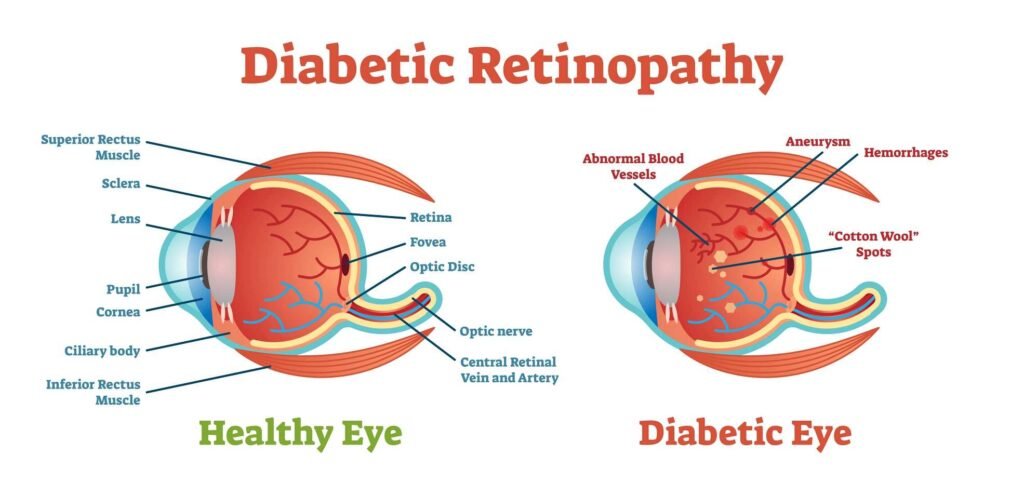
Diabetic retinopathy is a serious complication of diabetes caused by damage to the tiny blood vessels in the retina due to prolonged high blood sugar levels. Over time, this can lead to bleeding, swelling, or retinal detachment, resulting in severe vision loss if left untreated.
Digital retinal imaging offers a critical tool for diabetic retinopathy monitoring, enabling ophthalmologists to detect changes in blood vessel health during routine check-ups. Early detection allows for immediate action, such as:
- Adjusting blood sugar control
- Administering laser treatments to seal leaking vessels
- Using anti-VEGF injections to control abnormal vessel growth
Regular imaging ensures that any worsening condition is quickly identified, which prevents severe complications.
Cataracts

Though digital retinal imaging is not primarily intended for cataract diagnosis, it provides invaluable assistance in preparing for cataract surgery. Cataracts cloud the natural lens of the eye, obstructing light from passing through the retina, but imaging offers a clear look at the retina before the operation.
By assessing the retina’s health with high-resolution images, doctors can ensure there are no underlying retinal conditions-such as macular degeneration or diabetic retinopathy-that might complicate surgery or require additional treatment. This thorough evaluation ensures that surgery proceeds smoothly, maximizing outcomes and minimizing the risk of postoperative complications.
Enhancing Patient Experience With Digital Retinal Imaging
Beyond its diagnostic power, digital retinal imaging improves the overall patient experience in eye care. Traditional methods, such as pupil dilation, can be uncomfortable and time-consuming for patients, often resulting in blurred vision for hours after the exam.
In contrast, non-invasive eye scanning with digital retinal imaging is quick and painless, requiring no physical contact with the eye. This convenience does the following:
- Reduces patient anxiety
- Encourages routine exams
- Increases compliance with follow-up care
Another key benefit is the immediate availability of results. With real-time imaging, ophthalmologists can review scans with patients during the same visit, fostering transparent communication and better patient understanding.
Seeing the images of their retina empowers patients to grasp the seriousness of conditions like glaucoma or diabetic retinopathy, leading to improved adherence to prescribed treatments.
Additionally, digital retinal imaging allows eye care providers to document and share images with specialists remotely, facilitating seamless referrals and consultations. This capability is especially beneficial for patients in remote areas or those with limited access to specialty care. It enables timely second opinions and collaborative care, reducing delays in treatment.
Finally, integrating this technology enhances a clinic’s reputation. Patients value advanced, patient-friendly solutions and practices that offer optical coherence tomography (OCT) and digital retinal imaging, which are often seen as more professional and innovative, helping attract and retain patients over the long term.
The Importance of Long-Term Monitoring

Long-term monitoring through digital retinal imaging provides immense benefits, particularly for chronic conditions like glaucoma and diabetic retinopathy. High-resolution images are stored for future comparison, allowing ophthalmologists to track disease progression and adjust treatments accordingly. This capability is crucial because many eye diseases, such as macular degeneration, progress gradually and might not present symptoms until they are advanced.
By regularly updating retinal images, healthcare providers can detect even the smallest changes and intervene before significant vision loss occurs. Continuous monitoring enables more personalized and effective care, improving long-term outcomes for patients at risk of vision impairment.
Frequent imaging allows clinicians to identify subtle signs of worsening conditions and make timely decisions, whether to:
- Adjust medications
- Recommend surgical interventions
- Initiate new therapies
It also helps build a reliable baseline, which is especially valuable for high-risk patients with complex medical histories.
Moreover, monitoring through digital retinal imaging ensures consistency when patients switch providers or visit specialists. All historical data can be easily shared and accessed.
For ophthalmologists, it enhances the precision and confidence in treatment plans, fostering better trust and engagement with patients over time.
Risks and Limitations of Digital Retinal Imaging
While digital retinal imaging offers significant advantages, it’s essential to recognize its limitations. Not all eye conditions can be detected through retinal imaging alone.
For example, some diseases, such as cataracts or certain optic nerve disorders, may require additional diagnostic tests for a complete assessment.
Moreover, the availability of digital retinal imaging can be restricted by its high cost, making it less accessible in smaller clinics or regions with limited resources. In some cases, operator skill is required to ensure accurate results, which means the effectiveness of the imaging might depend on the technician’s expertise.
Invest in Digital Retinal Imaging to Elevate Your Practice
For physicians and clinics, investing in digital retinal imaging enhances diagnostic precision and supports superior patient outcomes through non-invasive eye scanning. Its ability to store high-resolution images enables long-term monitoring, helping providers track disease progression and optimize treatment plans.
With this technology, you position your practice at the forefront of eye care innovation, ensuring early detection, precise management, and better patient retention. Stay ahead of the curve-explore digital retinal imaging solutions today to offer cutting-edge care and maintain a competitive edge.
Shop now for the latest equipment at Nava Ophthalmic and discover how these tools can revolutionize your clinical practice.

Matthew Strachovsky, M.D.
Dr. Strachovsky's undergraduate training began in Boston, Massachusetts at Boston University and was completed at Stony Brook University in Long Island, NY. There he graduated Summa Cum Laude, obtaining a Bachelor of Science degree in Biology with special recognition for academic achievement.
He continued his education at Stony Brook School of Medicine and graduated with the additional designation of the "MD with Recognition" program. He worked as an intern in Internal Medicine at Winthrop University Hospital in NY and pursued a residency at Stony Brook University Hospital in Ophthalmology acting as Chief Resident in his final year. He completed his fellowship training in Vitreoretinal disease with a major emphasis on the diagnosis and management of retinal vascular diseases under the direction of Dr. Michael O'Brien at Koch Eye Associates in Rhode Island.
Dr. Strachovsky has presented research at the annual Association for Vision and Research in Ophthalmology meeting and published articles in journals including, Investigative Ophthalmology and Visual Science and The Journal of Neuro-ophthalmology.
Dr. Strachovsky's professional interests include the management of Age-Related Macular Degeneration and diabetic eye disease. He is Board Certified in Ophthalmology and a member of the American Academy of Ophthalmology, American Society of Retina Specialists, Young Ophthalmologist Network, and Leading Physicians of the World.
" I believe that the physician/patient relationship is more important than ever. Being an Ophthalmologist allows me to help patients and build a foundation of trust, knowledge, and professionalism when it comes to eye care".