Table of Contents
Comprehensive eye exams are essential to preventive health care for people of all ages. The American Academy of Ophthalmology suggests that adults get full eye exams regularly. These exams check vision, prescribe glasses, and look for signs of eye disease.
Ophthalmologists and optometrists use screenings to find early signs of eye disease. This helps prevent problems with the patient’s vision. Early detection allows patients to commence treatment quickly and prevent vision loss in the future.
Glaucoma screenings are a standard component of any comprehensive eye exam. Glaucoma is the leading cause of preventable blindness worldwide.
Glaucoma typically features increased intraocular pressure, which leads to progressive optic nerve damage. Without treatment, glaucoma causes vision loss and eventually blindness. Effective treatments are available and can reduce the risk of vision loss in people with glaucoma.
Tonometry is the preferred method for routine glaucoma monitoring. Eye care practices can choose between various tonometry procedures depending on which best serves their patients.
About Tonometry
Tonometry is any test that checks intraocular pressure (IOP). Doctors use tonometry screenings to measure the pressure in the eye’s anterior chamber, just behind the cornea. Too much fluid in the eye can raise eye pressure. This high pressure can be a sign of glaucoma.
High IOP can be a side effect of some medications. It can also result from eye injuries or health issues like diabetes or high blood pressure.
Identifying excessive IOP is critical for diagnosing and managing glaucoma and other ocular conditions. Tonometry is a safe, effective, and non-invasive method for checking IOP.
Tonometry tests use a calibrated instrument to measure intraocular pressure. Several methods exist for conducting tonometry screenings. All methods work well and cause little to no discomfort for patients.
 Types of Tonometry Tests
 Types of Tonometry Tests
Eye care practices can use various tonometry devices to check IOP. Investing in different types of technology can be helpful if not all patients respond to one device. It also proves helpful if researchers need more readings to confirm results.
Tonometry devices require regular maintenance, appropriate care, and calibration to ensure accurate results. Staff at eye care practices should know the full operating instructions for each tonometry device. This ensures it is set correctly for every patient. If you have questions about device operation, contact the manufacturer or customer service team for assistance.
- Applanation Tonometry: The word applanation means flattening and applanation tonometry involves gently flattening the front of the eye. Applanation tonometry devices have small, disk-shaped extensions that gently rest on the eye’s surface.
The device measures the pressure it takes to flatten the eye’s surface. Applanation is the most accurate type of tonometry. It can be used after other methods show possible IOP increases.
Before performing an application, providers give numbing eye drops and put a fluorescent dye in the eye. This helps to separate the tear film from the cornea.
The provider then places the application device on the eye’s surface to get an IOP reading. The test is painless and takes only a few seconds.
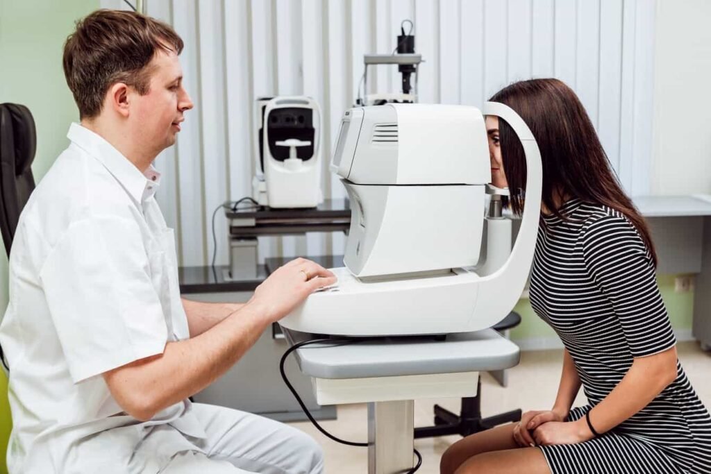
- Non-Contact (Air-Puff) Tonometry: Non-contact tonometry measures IOP by directing a puff of air at the cornea. The device measures how the air pressure changes the shape of the cornea as it pushes on the surface.
This type of test does not require numbing or dyeing the eyes. The patient sits in front of the device, places their chin in the chin rest, and leans their forehead into the forehead rest. The main drawback is the likelihood of patients reflexively closing their eyes as they feel the air.
You can repeat the test until you collect an accurate reading. This screening is painless and non-invasive.
- Rebound Tonometry: Rebound tonometry devices use a small plastic ball to touch the eye and check the pressure. These devices are handheld, and a stainless steel wire attaches the ball, which the electromagnetic field holds in place. When activated, the ball moves toward the eye until it contacts the cornea. The ball slows as it touches the eye, and researchers use that deceleration rate to calculate IOP.
This tonometry method is painless and does not require numbing drops or dyes. Some rebound tonometry devices are suitable for home use. Patients can use them to take regular IOP measures to identify changes over time.
- Indentation Tonometry: Indentation tonometry uses a small probe to touch the eye and measure the pressure. The probe checks the pressure inside the eye by measuring the force needed to make a small dent on the eye’s surface.
These can be handheld tonometers or tabletop devices. Tabletop indentation tonometers use a puff of air to bring the silicone-tipped probe into contact with the cornea.
Eye care professionals administer numbing drops before performing indentation tonometry. Patients should place their chin on the device’s chinrest and hold their eyes open to accommodate the probe. The test is painless and takes only seconds.
Administering Tonometry Screenings
Physician assistants, nurses, and eye doctors can administer tonometry screening. In some practices, support staff do routine screenings. This allows doctors to review the results before seeing the patient and discuss any findings.
During the screening, it is important to ensure patients are comfortable and cooperate with the instructions. Flinching or blinking when a tonometry device comes near the eye is common. Staff should be ready to repeat the process until they get an accurate reading patiently.
Staff can help patients relax and avoid blinking by offering a few helpful suggestions:
- Explain what patients can expect before beginning the test. This prevents any surprises and helps patients stay physically relaxed.
- Ask patients if they are sitting comfortably. They should not strain or reach to make contact with the chin rest on the tonometry device. Comfortable posture improves tonometry accuracy.
- Ask patients to focus on a spot behind the provider rather than looking at the tonometer tip. This helps them avoid blinking or flinching as the tip approaches their eye.
- Ask patients to keep both eyes wide open. Squeezing one eye shut can make it more challenging to test the open eye.
Interpreting Tonometry Results
IOP is measured in millimeters of Mercury (mmHg). While pressure varies from person to person, the typical range is between 12-22 mmHg. A measure above 20mmHg can indicate possible glaucoma.
 Tonometry is only one test for diagnosing glaucoma. Increased IOP does not constitute a definitive diagnosis. Instead, doctors should consider high intraocular pressure measurements in context with other screenings and the patient’s medical history.
If a patient shows increased IOP, eye care professionals will conduct additional screening to determine the cause. They might recommend a slit lamp exam to check the inner parts of the eye closely. They may also suggest imaging to look at the optic nerve for any signs of damage.
Repeated tonometry tests can help determine if a patient has pressure changes. These changes may relate to factors like the time of day, diet, or recent exercise.
Eye care professionals need to discuss medical history with patients, including current medications and any relevant health conditions. This helps providers check for other causes of high intraocular pressure, such as high blood pressure or medication side effects.
Benefits of Tonometry
The available devices for in-office tonometry are noninvasive, quick, and effective methods for monitoring eye pressure. They can provide accurate measurements that indicate whether intraocular pressure is within normal ranges. Accurate screening for early signs of glaucoma is important because it’s the best way to detect the condition early.
Glaucoma doesn’t usually cause pain or discomfort. The damage to the optical nerve happens gradually and doesn’t lead to immediate vision changes. If glaucoma gets worse, it can damage the optic nerve. This affects side vision and can lead to loss of central vision.
Tonometry identifies the first signs of glaucoma before symptoms arise. Patients can start glaucoma treatment before they notice any vision changes. This may help completely prevent future vision loss.
Treatments for glaucoma include:
- Prescription eye drops: Prescription eye drops can reduce intraocular pressure and improve fluid drainage in the eyes.
- Medications: Oral medications may reduce IOP to prevent damage to the optic nerve.
- Laser treatments: Minimally invasive laser treatments can dramatically improve eye drainage.
- Surgery: Surgery to place a stent or increase the natural drainage can improve IOP.
Tonometry is a standard part of comprehensive eye care. Measuring intraocular pressure is one way of assessing eye health and identifying early signs of glaucoma. Early diagnosis and treatment can prevent vision changes and give patients a future without the threat of vision loss.
Eye care providers can choose the best tonometry technologies for their practices. Selecting a tonometry device or multiple devices depends on patient population, office layout, and staff expertise.
If you have questions about which tonometry devices are best for your practice, contact the team at Shape Ophthalmics. We can help you decide which device is the right investment.
You can also learn more about tonometry by visiting these websites:
- WebMD: Tonometry: Purpose, Procedure, and Results
- Cleveland Clinic: Tonometry: What Is It, Types, Test Procedure & Results
- American Academy of Ophthalmology: IOP and Tonometry – EyeWiki
- StatPearls: Tonometry – StatPearls – NCBI Bookshelf
Matthew Strachovsky, M.D.
Dr. Strachovsky's undergraduate training began in Boston, Massachusetts at Boston University and was completed at Stony Brook University in Long Island, NY. There he graduated Summa Cum Laude, obtaining a Bachelor of Science degree in Biology with special recognition for academic achievement.
He continued his education at Stony Brook School of Medicine and graduated with the additional designation of the "MD with Recognition" program. He worked as an intern in Internal Medicine at Winthrop University Hospital in NY and pursued a residency at Stony Brook University Hospital in Ophthalmology acting as Chief Resident in his final year. He completed his fellowship training in Vitreoretinal disease with a major emphasis on the diagnosis and management of retinal vascular diseases under the direction of Dr. Michael O'Brien at Koch Eye Associates in Rhode Island.
Dr. Strachovsky has presented research at the annual Association for Vision and Research in Ophthalmology meeting and published articles in journals including, Investigative Ophthalmology and Visual Science and The Journal of Neuro-ophthalmology.
Dr. Strachovsky's professional interests include the management of Age-Related Macular Degeneration and diabetic eye disease. He is Board Certified in Ophthalmology and a member of the American Academy of Ophthalmology, American Society of Retina Specialists, Young Ophthalmologist Network, and Leading Physicians of the World.
" I believe that the physician/patient relationship is more important than ever. Being an Ophthalmologist allows me to help patients and build a foundation of trust, knowledge, and professionalism when it comes to eye care".