Table of Contents
The CDC says about 9.6 million people in the United States had diabetic retinopathy in 2021. This staggering number underscores the need for regular eye examinations and advanced diagnostic tools to prevent irreversible vision loss.
Eye health is vital to overall well-being, affecting everything from daily tasks to long-term quality of life. With an increasing number of individuals experiencing vision-related issues, regular eye exams have become crucial to preventive healthcare.
Digital eye exams represent a groundbreaking advancement. It allows eye care professionals to diagnose and monitor conditions with greater accuracy.
Discover how these advanced exams work, their benefits, and why they are game-changers in modern eye care.
The Rise of Digital Eye Exams
Traditional eye exams have long been the standard for detecting refractive errors, eye diseases, and overall vision health. However, eye exam technology has evolved significantly. It offers more precise, automated, and comprehensive evaluations.
Digital eye exams use advanced imaging, artificial intelligence (AI), and noninvasive scanning. These tools give better insights into eye health and are essential for today’s eye care in ophthalmology and optometry.
What Is a Digital Eye Exam?
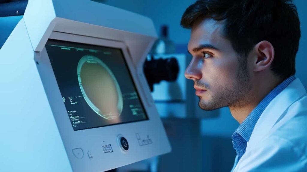
A digital eye exam is a modern test that checks eye health. It uses new technology to be more accurate than old methods. These exams leverage cutting-edge imaging and artificial intelligence to detect potential vision issues before symptoms appear.
Digital eye exams are different from regular exams. They do not depend much on manual methods or patient opinions. Instead, they use automated tools to provide accurate, data-based information.
Key Components of a Digital Eye Exam
Modern eye care advancements have transformed how professionals diagnose and treat vision-related conditions. Integrating digital technologies has improved the accuracy and efficiency of eye examinations.
A digital eye exam uses advanced technology to provide a complete eye health check.
Retinal Imaging
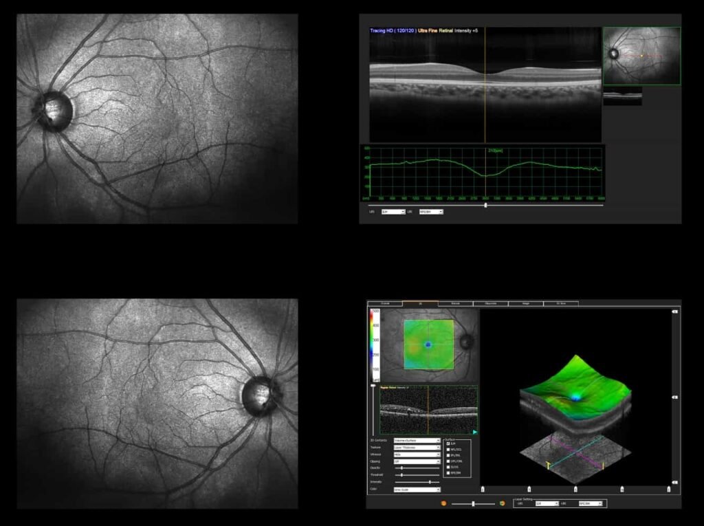
Retinal imaging captures high-resolution retina images, enabling early detection of eye diseases such as glaucoma, macular degeneration, and diabetic retinopathy. By providing detailed images, retinal imaging helps ophthalmologists monitor changes over time and detect abnormalities before symptoms appear, improving the chances of early intervention.
Automated Refraction
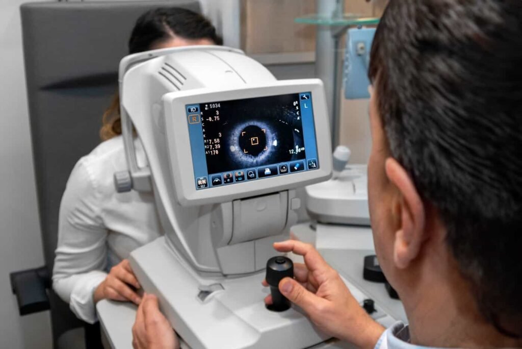
Determines the exact prescription for glasses or contact lenses by analyzing how light passes through the eye. This technology minimizes human error and provides highly accurate lens prescriptions, reducing the need for manual refraction tests.
Corneal Topography
It maps the shape of the cornea to detect irregularities that may cause vision problems. This is useful for diagnosing conditions like keratoconus and assessing the fit of specialty contact lenses.
Optical Coherence Tomography (OCT)
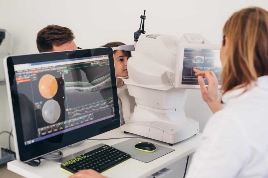
This noninvasive imaging test provides detailed retina and optic nerve cross-sectional images. OCT helps find diseases like glaucoma, macular degeneration, and diabetic retinopathy. It shows tiny changes in retinal tissue that standard exams might miss.
AI-Powered Analysis
Some systems incorporate artificial intelligence to detect patterns indicative of eye diseases faster and more accurately than human analysts alone. AI enhances diagnostic precision by analyzing vast amounts of data and identifying conditions at earlier stages than traditional examination methods.
These components ensure that digital eye exams provide a detailed and comprehensive view of a patient’s eye health. They enable early intervention and more effective treatments.
The Benefits of Eye Exams Using Digital Technology
The transition from conventional eye exams to digital eye exams brings numerous advantages. This is why you should choose digital eye exams over traditional methods.
Enhanced Accuracy
Digital eye exams minimize human error by using precise automated measurements. These computerized systems evaluate the eye’s refractive errors and structural integrity with high accuracy, reducing variability caused by human assessment.
Digital eye exams give reliable results. They help create more accurate prescriptions for glasses or contacts and provide a complete examination of the eye’s structure.
Furthermore, integrating digital technology allows for better customization of treatment plans. This ensures that each patient receives care tailored to their specific visual needs.
Early Detection of Eye Diseases
Conditions such as diabetic retinopathy, glaucoma, and macular degeneration often develop without noticeable symptoms. Digital eye exams using clear images and AI analysis help eye care professionals find these conditions early.
Digital exams help catch problems early, allowing for better management of diseases that could cause permanent vision loss. They do this by capturing and saving detailed retinal scans over time.
Additionally, these exams help identify subtle changes in eye health. The ophthalmologists implement treatment strategies that preserve long-term vision before significant damage occurs.
Faster and More Efficient Process
Automated systems speed up the eye exam process, reducing patients’ time in the clinic while providing more comprehensive results.
Digital eye exams make the whole process easier. They capture images and analyze data quickly, helping patients get a complete evaluation in less time.
This efficiency is particularly beneficial for high-volume clinics. It allows them to serve more patients without compromising on quality.
Additionally, digital records enable instant access to patient history. They eliminate the need for redundant tests and improve workflow.
Better Patient Education
Digital eye exams show clear images of eye health issues. This helps eye care professionals explain diagnoses and treatment plans to patients. High-resolution imaging allows patients to see detailed scans of their eyes, helping them understand their condition better. This visual approach improves communication between doctors and patients and encourages adherence to treatment plans.
When patients understand the importance of their eye health through clear images, they are more likely to act to manage their vision.
Comprehensive Data Storage
Unlike paper records, digital imaging and test results can be stored electronically, allowing easy access to historical data to monitor changes over time. This digital record-keeping system improves efficiency and helps eye care professionals accurately track disease progress and treatment success.
Furthermore, it reduces the risk of misplaced files and allows seamless integration with other diagnostic tools, ensuring a holistic approach to patient care.
Clinicians can quickly access past scans and test results, which helps them make better decisions and improves patients’ outcomes. It also smooths workflows in busy eye care practices.
How Digital Eye Exams Work
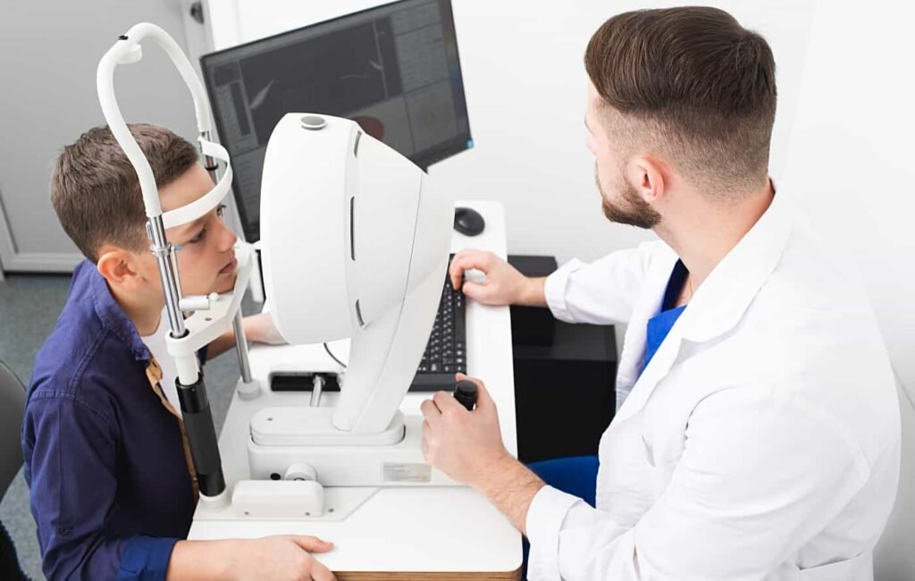
To ensure patients receive the most comprehensive evaluation, digital eye exams follow a systematic approach that leverages advanced technology. A digital eye exam follows a structured process to provide a thorough assessment.
Below, we take a closer look at the step-by-step eye exam process.
1. Patient History and Symptoms Review
Eye care professionals ask about medical history, family history, and vision concerns. This step helps identify underlying conditions such as diabetes, hypertension, or genetic predispositions affecting eye health. A thorough review enables a more targeted examination and ensures that we carefully monitor potential risk factors.
2. Preliminary Tests
- Preliminary tests include depth perception, color vision, and eye muscle movement evaluations. These tests give essential information about how the eyes work together. They can also find problems that may show issues like Amblyopia
- Strabismus
- Optic nerve disorders
By identifying potential problems early, eye care professionals can recommend appropriate interventions to preserve vision quality.
3. Automated Refraction
Automated refraction determines the precise prescription using digital instruments. This method enhances accuracy by analyzing how light is bent as it enters the eye. It provides a more refined and personalized prescription compared to traditional methods.
Digital refraction minimizes human error and ensures precise adjustments for optimal vision correction.
4. Digital Retinal Imaging
Digital retinal imaging captures high-resolution images of the retina and optic nerve. This advanced imaging technology enables early detection of diseases such as:
- Glaucoma
- Macular degeneration
- Diabetic retinopathy
This is done by offering a detailed view of the eye’s internal structures. Ophthalmologists can track changes by storing and comparing retinal images over time, which helps them provide better treatments and improve patient outcomes in the long run.
5. OCT Scanning
This provides detailed cross-sectional images to detect underlying diseases. OCT scanning uses light waves to take layered images of the retina. This helps identify conditions like macular degeneration, diabetic retinopathy, and glaucoma earlier than traditional methods.
This non-invasive procedure offers a high-resolution view of the eye’s internal structures, aiding in precise diagnoses and better treatment planning.
6. Comprehensive Analyzing
The data is analyzed using AI-powered software for a detailed report on eye health. Advanced machine learning algorithms evaluate these scans to detect even the slightest abnormalities. This ensures that we overlook no early signs of disease.
The AI-driven method makes the diagnostic process easier. It helps doctors by reducing their workload. At the same time, it improves accuracy and consistency in patient assessments.
7. Final Consultation
The eye care professional thoroughly reviews the examination results and explains the findings using digital imaging and AI-assisted techniques.
Also, they provide personalized recommendations tailored to the patient’s needs, which may include the following:
- Corrective lenses
- Prescription adjustments
- Treatment options for detected conditions
- Additional diagnostic tests
This step helps patients understand their eye health and how to maintain or improve their vision.
Understanding the Role of Eye Exam Technology in Modern Eye Care
Technology has revolutionized eye exam procedures. Below, we explore some of the most influential advancements in eye exam technology.
Artificial Intelligence (AI) in Eye Exams
AI enhances diagnostic capabilities by identifying diseases through pattern recognition in retinal images. AI tools can analyze thousands of retinal scans. They can find early signs of conditions like diabetic retinopathy and glaucoma. This happens before symptoms appear.
These advanced systems help reduce misdiagnoses and improve treatment timelines, ensuring patients receive on-time and accurate care.
Telemedicine for Eye Care
Remote eye exams and consultations allow patients to receive expert evaluations without visiting a clinic. This technology is especially beneficial for individuals in remote or underserved areas, providing them with access to high-quality eye care from specialists.
Eye care professionals can effectively diagnose and manage conditions through video consultations and digital imaging. This bridges the gap between accessibility and quality healthcare.
Portable Digital Retinal Imaging
Handheld imaging devices enable mobile screenings in rural or underserved areas. These portable devices allow healthcare providers to conduct eye exams outside traditional clinic settings, making detecting and diagnosing eye diseases possible in populations with limited access to specialized care.
These tools use high-resolution images and advanced software. They give quick insights into a patient’s retinal health, helping improve access to eye care worldwide.
Smart Contact Lenses
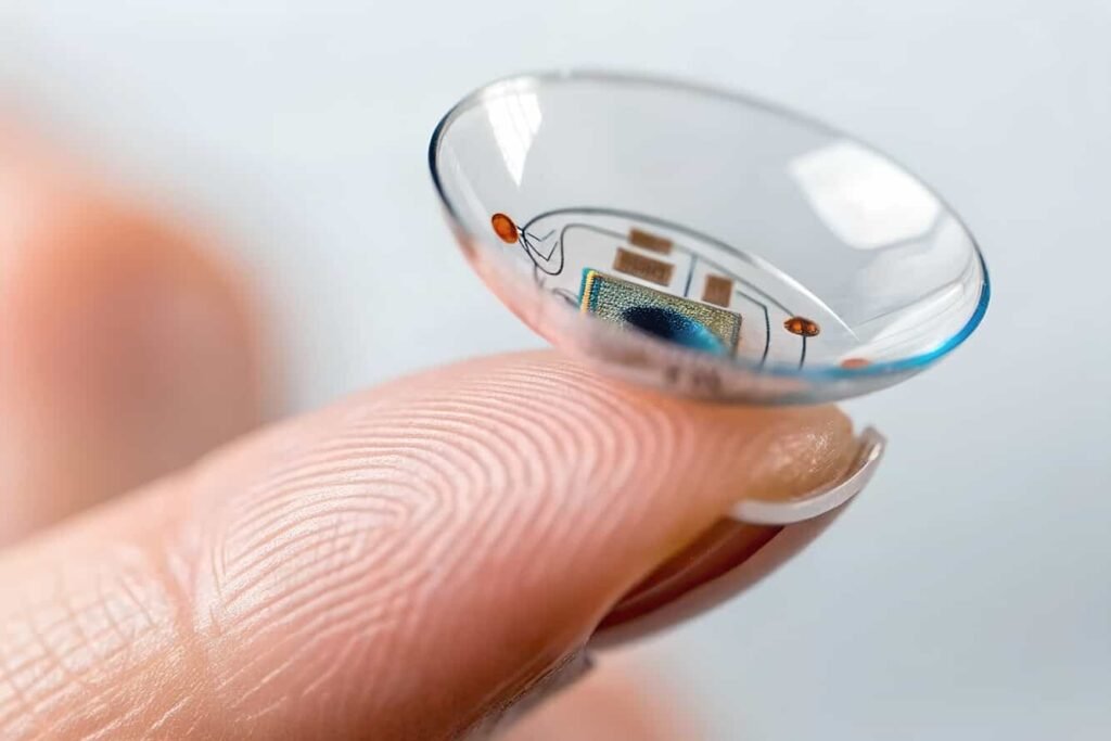
This is an emerging technology that could monitor intraocular pressure for glaucoma patients. These new lenses have small sensors that measure pressure changes in the eye. They send data to a connected device for real-time monitoring.
The noninvasive approach efficiently tracks glaucoma progression for patients and doctors. It also reduces the need for frequent in-office visits while enhancing early detection of dangerous pressure fluctuations.
The integration of these technologies continues to enhance the accuracy, efficiency, and accessibility of eye care.
How Often Should You Get a Digital Eye Exam?
Regular digital eye exams are vital for maintaining good vision health. The recommended frequency varies based on age and risk factors:
- Adults (18-60 years): Every two years if no underlying conditions exist
- Adults over 60: Annually, because of increased risk of eye diseases.
- Individuals with risk factors: Annually or as an eye care professional recommends.
People with diabetes, high blood pressure, or a family history of eye diseases may need more exams.
Why Your Practice Needs Digital Eye Exams
Digital eye exams are not just a technological advancement. They are a necessity in modern eye care. Their ability to detect diseases early, improve diagnostic accuracy, and enhance patient education makes them indispensable for any practice.
At Nava Ophthalmic, we understand the importance of high-quality, affordable ophthalmic products. Our commitment to providing the largest selection ensures your practice has the best tools available. We prioritize quality, reliability, and service, proving our dedication through the products and support we offer.
Explore our extensive digital eye exam equipment catalog, or contact our expert team for personalized assistance.

Matthew Strachovsky, M.D.
Dr. Strachovsky's undergraduate training began in Boston, Massachusetts at Boston University and was completed at Stony Brook University in Long Island, NY. There he graduated Summa Cum Laude, obtaining a Bachelor of Science degree in Biology with special recognition for academic achievement.
He continued his education at Stony Brook School of Medicine and graduated with the additional designation of the "MD with Recognition" program. He worked as an intern in Internal Medicine at Winthrop University Hospital in NY and pursued a residency at Stony Brook University Hospital in Ophthalmology acting as Chief Resident in his final year. He completed his fellowship training in Vitreoretinal disease with a major emphasis on the diagnosis and management of retinal vascular diseases under the direction of Dr. Michael O'Brien at Koch Eye Associates in Rhode Island.
Dr. Strachovsky has presented research at the annual Association for Vision and Research in Ophthalmology meeting and published articles in journals including, Investigative Ophthalmology and Visual Science and The Journal of Neuro-ophthalmology.
Dr. Strachovsky's professional interests include the management of Age-Related Macular Degeneration and diabetic eye disease. He is Board Certified in Ophthalmology and a member of the American Academy of Ophthalmology, American Society of Retina Specialists, Young Ophthalmologist Network, and Leading Physicians of the World.
" I believe that the physician/patient relationship is more important than ever. Being an Ophthalmologist allows me to help patients and build a foundation of trust, knowledge, and professionalism when it comes to eye care".
